Hematopoietic growth factor inducible neurokinin-1 gene

a hematopoietic growth factor and neurokinin technology, applied in the field of immunology, can solve the problems of significant reduction of normal blood cells in circulation, stem cells produced uncontrollably, and the inability to produce stem cells, and achieve the effect of reducing side effects

Inactive Publication Date: 2005-09-06
RUTGERS THE STATE UNIV
View PDF0 Cites 13 Cited by
  • Summary
  • Abstract
  • Description
  • Claims
  • Application Information

AI Technical Summary

Benefits of technology

[0034]In a more specific embodiment, the invention relates to methods for using such polynucleotides, polypeptides and antibodies for preventing or treating acute and chronic myeloid leukemia and acute and chronic lymphocytic leukemia, as well as the B-cell subtype of Hodgkin's and non-Hodgkin's lymphomas. More specifically, for example, the compositions of the present invention may be used for the treatment of Acute Myelocytic Leukemia, which is associated with the accumulation of immature blast cells, wherein the administration of HGFIN compositions may enhance the maturation of the affected cells thus alleviating the leukemic condition and the anemia and low platelet blood count associated with this disease. Further, the compositions of the present invention may also be useful in the treatment of Acute Lymphocytic Leukemia which is associated with increased proliferation of immature lymphocytes, wherein the administration of an HGFIN composition may inhibit and or slow down proliferation and promote differentiation, helping the cells mature before becoming the cells of the peripheral blood system.
[0035]The compositions of the present invention may also be useful in the treatment of Chronic Myelogenous Leukemia which is marked by the abnormal proliferation of immature granulocytes in the BM and blood, wherein the administration of an HGFIN composition that includes an HGFIN antisense sequence or HGFIN immunospecific antibody may inhibit and or slow down proliferation, allowing the developing cells time to mature before differentiating into the cells of the peripheral blood system. Further, the compositions of the present invention may also be useful in the treatment of Chronic Lymphocytic Leukemia which is marked by mature but poorly functioning lymphocytes circulating in the blood, wherein the administration of an HGFIN composition may inhibit and or slow down the earlier stages of proliferation, allowing more time for the cells to mature before terminal differentiation.
[0037]In another embodiment HGFIN immunospecific antibodies may be used to target disease cells, these antibodies may also be conjugated with chemo- or radio-toxic agents to kill off leukemia or lymphoma associated cells. Such a method would also allow for the reduction of side effects caused by the administration of such cyto- or radio-toxic elements by reducing the amount of dosage of the toxic agent needed to kill affected cells.

Problems solved by technology

However, in the throes of a diseased state, the BM may not produce enough stem cells, may produce too many stem cells or various ones produced may begin to proliferate uncontrollably.
Further complications arise when these stem cells or their associated progenitor are not able to differentiate into the various morphologically recognizable and functionally capable cells circulating in the blood.
These cells are unable to mature and differentiate properly leading to a significant reduction of normal blood cells in the circulation.
The accumulation of blast cells in the BM prevents production of other cell types resulting in anemia and low platelet blood counts.
Traditional methods of treating these B-cell malignancies, which includes chemotherapy and radiotherapy, have limited utility due to toxic side effects.
Short-term side effects of chemotherapy may include significant toxicity, extreme nausea, vomiting, and serious discomfort.
The short-term side effects of radiotherapy may include extreme nausea, vomiting, serious discomfort, sterility and infertility.
These side effects may be moderated by reduced dosages, however, that increases the risk of remission.
However, these procedures are plagued with exorbitant cost and high rates of failure.
It is both difficult and costly to locate a sufficient donor and even when one is located, rejection of the transplanted cells often takes place, which in turn can lead to graft versus host disease.
Most often, these treatments also include a combination of both chemo and radiotherapies, hence, the concomitant risks involved therein would apply here as well.

Method used


Image

  • Hematopoietic growth factor inducible neurokinin-1 gene
  • Hematopoietic growth factor inducible neurokinin-1 gene
  • Hematopoietic growth factor inducible neurokinin-1 gene

Examples


examples

[0189]The following description sets forth the general procedures involved in practicing the present invention. To the extent that specific materials are mentioned, it is merely for purposes of illustration and is not intended to limit the invention.

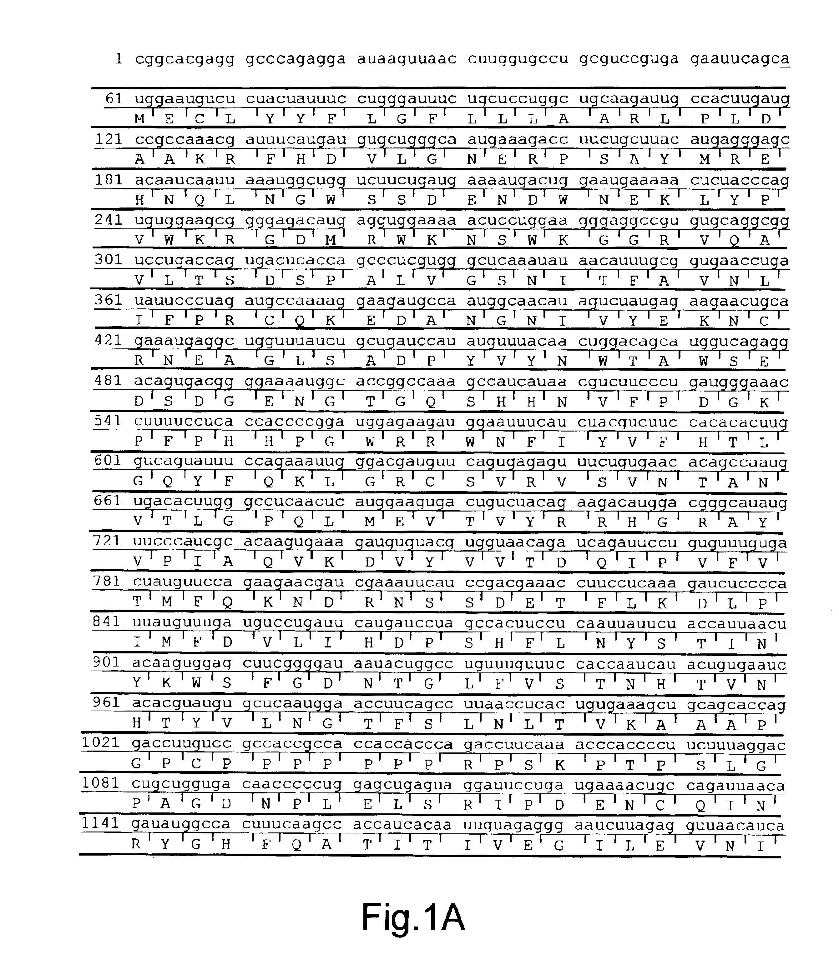
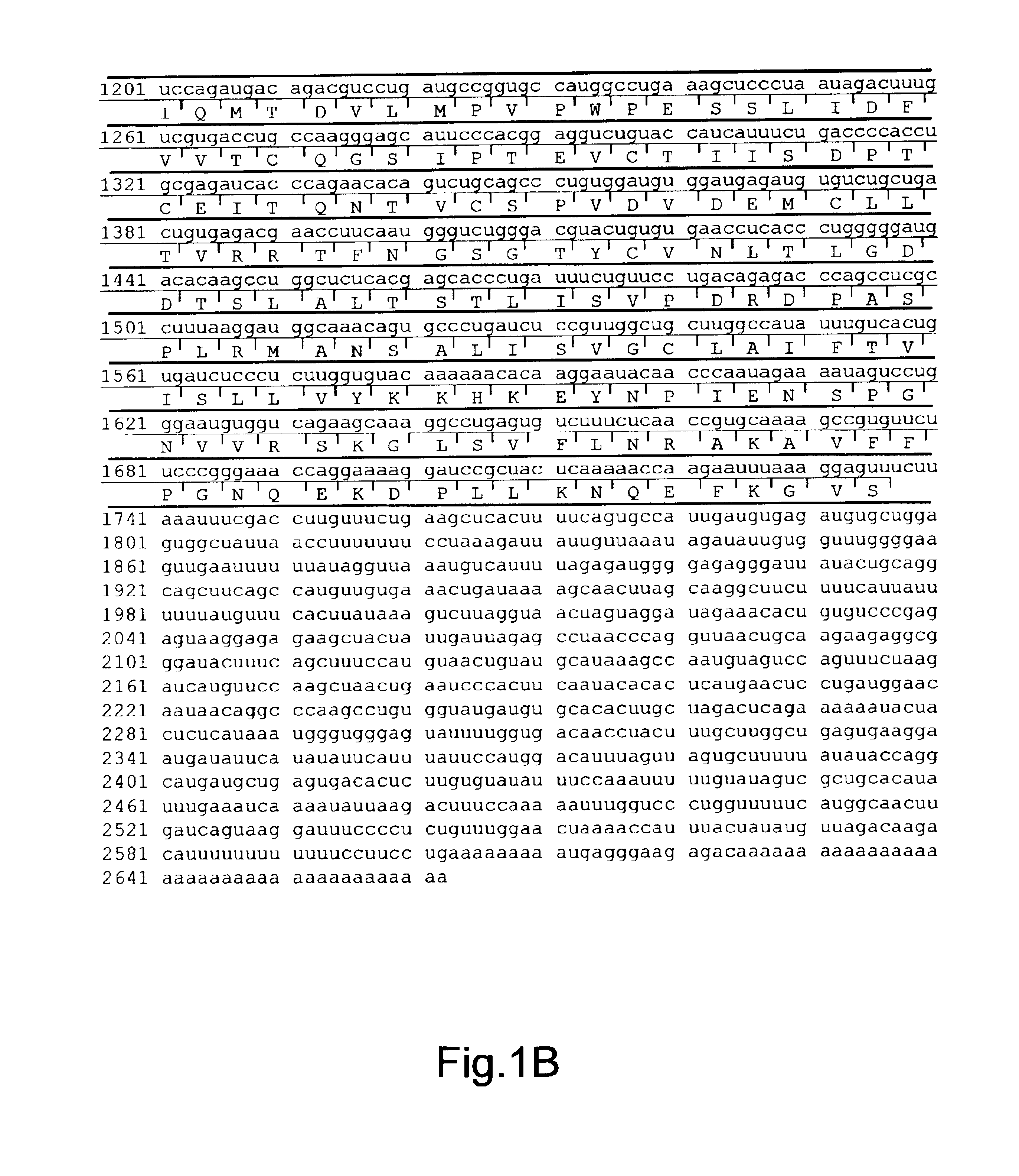
[0190]All ligands for the putative HGFIN transmembrane protein are unknown. Since the HGFIN clone was retrieved through screening of cDNA libraries with an NK-1-specific probe, it is believed that the natural ligand for NK-1 is likely responsible for interacting with HGFIN. The 3-D structures of the PKD region from HGFIN (FIG. 3A) was used to determine interactions with SP, which is the preferred ligand for NK-1 (FIG. 3C). Based on the putative spatial arrangement of the HGFIN protein (FIG. 2), SP could contact the extracellular PKD region, after all, the electrostatic differences between SP (17) and PKD could explain the formation of a possible complex (FIG. 3Q). See the examples below for further details.

[0191]Unless other-wise specifi...

example i

[0192]A. Reagents

[0193]Hoffman-La Roche (Nutley, N.J.) provided recombinant human (rh) IL-1α. Stem cell factor (rhSCF), rhIL-6, rhIL-11 and alkaline phosphatase (Alk Phos)-conjugated goat anti-rabbit IgG were purchased from R&D Systems (Minneapolis, Minn.). IL-1β and nerve growth factor (NGF) were purchased from Collaborative Research (Bedford, Mass.) and Amersham Life Science (Cleveland, Ohio) respectively. The following was purchased from Sigma (St Louis, Mo.): Isopropyl-D-Thioglactopyranoside (IPTG), SP, Ficoll Hypaque, lipopolysaccharide (LPS), Fibronectin-Fragment III-C (FN-IIIC), 12-0-tetradecanoylphorbol diester (TPA), dimethylsulfoxide (DMSO) and cytochemical staining kits for 2-naphthyl-acetate esterase and naphthol AS-D chloroacetate esterase. SP was dissolved in sterile distilled water and then immediately solublized with nitrogen gas. The reconstituted SP was used within two days. The immunology department of Genetics Institute (Cambridge, Mass.) provided the human G-CSF...

example 2

[0201]A. Cell Differentiation

[0202]Cell differentiation was performed with a myelomonocytic cell line, HL-60, or BM mononuclear cells (BMNC). HL-60 cells were chemically differentiated with TPA and DMSO for monocytes and granulocytes respectively (18, 19). BMNC were isolated from BM aspirate of healthy donors using Ficoll Hypaque density gradient. The IRB of UMDNJ-Newark Campus approved the use of human BM aspirate for these studies.

[0203]Further, BMNC cells were differentiated with M-CSF or G-CSF (500 U / ml for each) to monocytes and granulocytes respectively. Undifferentiated cells were cultured in parallel with only media. Culture media were replaced at two-day intervals until cytochemical staining determined that >90% of the cells were differentiated. At this time, cell differentiation was terminated and the cells analyzed by northern analyses for Id2 and HGFIN mRNA, and by immunoblot for Id2 protein. For HL-60 cultures, cytochemical staining was performed after three days with 1...


PUM

PropertyMeasurementUnit
pHaaaaaaaaaa
Tmaaaaaaaaaa
temperatureaaaaaaaaaa

Abstract

Bone marrow (BM) is the major organ where immune cells are derived. Homeostasis in the BM is maintained by inter- and intra-cellular interactions by the various subsets of BM cells. The present invention discloses the cloning of a new cDNA from stimulated BM stromal cells that was retrieved with a probe specific for the neurokinin-1 (NK-1) receptor. The cloned cDNA was designated ‘Hematopoietic Growth Factor Inducible Neurokinin-1 type’ (HGFIN) gene based on its expression in differentiated hematopoietic cells. Hence, the present invention provides a novel gene, HGFIN, which encodes a protein receptor that is involved in the regulation of hematopoietic proliferation and differentiation. The protein of the present invention may be involved as a central mediator of white blood cell, progenitor, differentiation, and therefore, may be useful in the prevention and treatment of lymphoproliferative syndromes such as B-cell related maladies, including but not limited to acute and chronic myeloid and lymphocytic leukemia as well as the B-cell subtype of Hodgkin's and non-Hodgkin's lymphomas.

Description

[0001]The present utility patent application claims priority to provisional patent application U.S. Ser. No. 60 / 241,881 (Rameshwar et al.), filed Oct. 20, 2000, the disclosure of which is incorporated by reference in its entirety herein.GOVERNMENT INTEREST[0002]This invention was made with government support by the following Public Health Service grants: HL-54973 and HL-57675 from the National Institute of Health and CA89868 from the National Cancer Institute. The government may own certain rights in the present invention.FIELD OF THE INVENTION[0003]The present invention relates to the field of molecular biology, immunology, and the regulation of lymphocytic cell proliferation and differentiation. In particular, this invention provides a novel gene, Hematopoietic Growth Factor Inducible Neurokinin-1 Gene (HGFIN-1), which was isolated from stimulated human Bone Marrow stromal cells. HGFIN-1 plays an important role in inducing white blood cell differentiation and may play a role in in...

Claims


Application Information

Patent Timeline
Patent Type & Authority Patents(United States)
IPC IPC(8): A61K47/48C07K14/435C07K14/72
CPCA61K47/48269C07K14/723A61K47/642A61P35/00
Inventor RAMESHWAR, PRANELA
Owner RUTGERS THE STATE UNIV
Why Eureka
Try Eureka