Quality control for cytochemical assays

a cytochemical and assay technology, applied in the field of quality control of cytochemical assays, can solve the problems of insufficient type of information available from such stains for accurate diagnosis, lack of inter-laboratory standardization, and inability to develop simple methods for accurately and reliably monitoring quality control

Inactive Publication Date: 2006-03-14
LAB VISION CORP
View PDF67 Cites 14 Cited by
  • Summary
  • Abstract
  • Description
  • Claims
  • Application Information

AI Technical Summary

Benefits of technology

[0028]In another embodiment of the present invention, the antigens can be attached, or affixed, directly onto a planar test platform, e.g., a glass microscope slide. This alternative embodiment eliminates the need for a separate matrix that adheres to the test platform with an adhesive backing. One or more antigen spots can be directly attached onto an area, or section, of the glass slide, leaving an appropriately large area for placement of the biological sample. In one embodiment of the present invention, the antigen is covalently attached, or coupled, to the test platform. A variety of covalent coupling chemistries are described

Problems solved by technology

Since IHC assays are relatively new in medical practice, simple methods for accurately and reliably monitoring quality control has not yet been developed.
Often, the type of information available from such stains is insufficient for accurate diagnosis.
With laboratories generating their own control tissues, there is little inter-laboratory standardization.
Standardized positive controls have been harder to achieve.
The use of multi-tumor tissue blocks as positive controls does not solve three important problems.
It is impossible to perform this type of analysis using tissue sections as controls since there is no practical method for quantitation and titration of antigen in a tissue section.
In addition, tissue sections as controls do not control for performance error.
Cutting (with a microtome) and mounting tissue sections on glass slides is labor intensive.
Because of the associated labor costs, few laboratories place a positive tissue control on each microscope slide.
Thus, if there is an error by placing an incorrect antib

Method used


Image

  • Quality control for cytochemical assays
  • Quality control for cytochemical assays
  • Quality control for cytochemical assays

Examples


example 1

Select Phage That Mimic Target Epitopes from Monoclonal Antibodies Commonly Used in Clinical Immunohistochemical Laboratories

[0088]For the device and methods describe herein, it is desirable to identify peptides that will mimic the antigen-specific interaction of antibody with the native antigen. These peptides will be suitable for use on either the quality control adhesive strips or glass slides described herein. Suitable peptide sequences identified as described herein are more amenable to consistent manufacture than the native protein itself. The monoclonal antibody clones suitable for use as described herein are those that recognize the antigen even after the tissue has been fixed with formalin and embedded in paraffin wax. These monoclonal antibody clones represent some of the most commonly used primary antibodies in the clinical IHC laboratory. The cyclic peptide libraries comprise a random assortment of peptides that have two cysteine residues that are linked by a disulfide. ...

example 2

Peptide (Antigen Mimic)—Matrix Optimization

[0102](a) Overview

[0103]Once an appropriate peptide sequence is identified, the next step is to determine the linear range of the peptide concentration on the control strips. In other words, how much peptide should be placed onto the nylon strip so as to be most sensitive to early reagent failure!

[0104]Carboxy-derivatized nylon or PVDF matrices allow high capacity covalent immobilization of proteins to the surface of the matrix while retaining biological activity. High capacity nitrocellulose membranes also serve to immobilize proteins or peptides in a manner adaptable to this invention. Processing steps include the covalent or non-covalent binding of protein to the membrane and a blocking step to quench the remaining covalent binding capacity, as described herein. Standardization is more readily achieved with proteins that are covalently, rather than non-covalently, adsorbed to matrices. Other desirable characteristics of nylon or PVDF mem...

example 3

Validation of Synthetic Control Strips

[0111]Each peptide mimic or antigen comprises a somewhat unique and independent product. Therefore, each needs to be independently validated. This section describes the process of validating the synthetic control strips to verify that (a) each synthetic control strip has a high signal to noise ratio, (b) the synthetic control strip is antibody-specific, and (c) each synthetic control strip is capable of detecting early reagent failure.

[0112](a) Measurement of signal to noise ratio

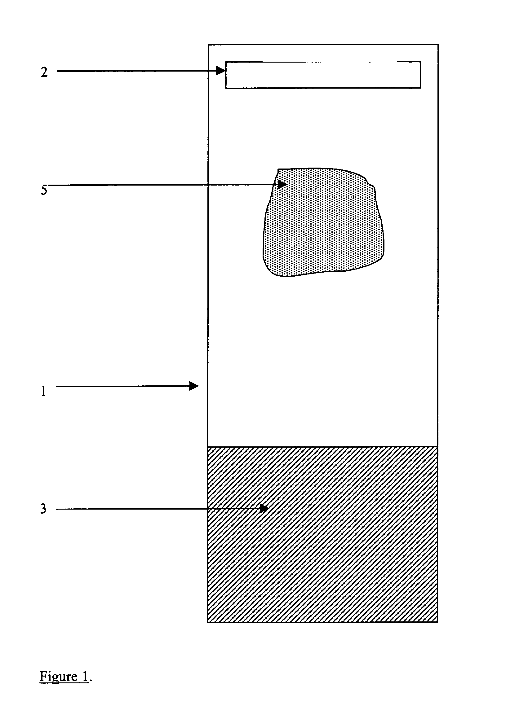
[0113]The synthetic control strip contains two distinct area: (1) the reagent surface that is devoid of bound peptide, and (2) the test area with specific peptide. The test strip containing no peptide should be used as a test for non-specific binding of antibody to the membrane itself. Excessive background to the negative control (surface without peptide) indicates the need to investigate the blocking procedure, buffer composition, and / or the washing procedure during th...


PUM

PropertyMeasurementUnit
Concentrationaaaaaaaaaa
Transparencyaaaaaaaaaa

Abstract

The invention describes quality control devices for assays that measure analytes in cells and tissue samples, and methods of use thereof. In particular, the quality control device comprises a matrix affixed with synthetic controls in different concentrations, or different synthetic controls. The quality control device can be adhered to a microscope slide via an adhesive or chemically attached to a microscope slide and processed simultaneously with a tissue sample.

Description

[0001]This application is a Continuation-in-Part of U.S. Application Ser. No. 09 / 291,351, filed Apr. 14, 1999, now U.S. Pat. No. 6,281,004, issued on Aug. 28, 2001, the entire teachings of which are incorporated herein by reference.GOVERNMENT SUPPORT[0002]The invention was supported, in whole or in part, by a grant 1R43CA81950-01 from The National Institute of Health. The Government has certain rights in the invention.BACKGROUND OF THE INVENTION[0003]During the past decade, immunohistochemical (IHC) stains have become an integral part of the diagnostic process in surgical pathology. IHC stains are used with conventional histopathological stains, as adjunctive assays. They are critical in correctly diagnosing poorly differentiated malignancies, viral infections, and tumor prognoses, such as in the use of estrogen receptor (ER) analysis for breast carcinoma. Since IHC assays are relatively new in medical practice, simple methods for accurately and reliably monitoring quality control h...

Claims


Application Information

Patent Timeline
IPC IPC(8): C12Q1/00G01N33/53G01N21/93G01N33/543
CPCG01N33/54386G01N21/93G01N2333/723Y10T436/105831Y10T436/106664Y10T436/107497Y10T436/25375Y10T436/2525Y10T436/25G01N1/312G02B21/34
Inventor SOMPURAM, SESHI R.RAMANATHAN, HALASYABOGEN, STEVEN A.
Owner LAB VISION CORP
Why Eureka
Try Eureka