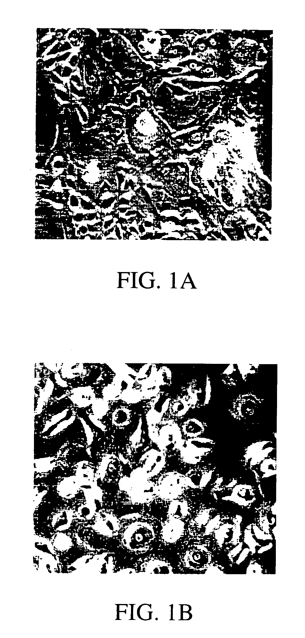
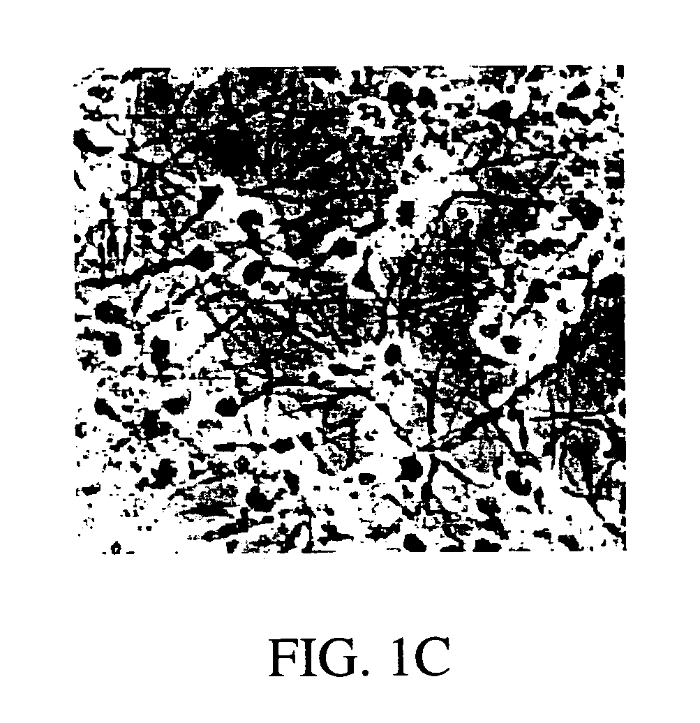
Transdiffentiation of transfected epidermal basal cells into neural progenitor cells, neuronal cells and/or glial cells
a technology of epidermal basal cells and transfected epidermal cells, which is applied in the field of neuronal tissue regeneration, can solve the problems of preventing the regeneration of neural tissues, hindering the development of neurological injury therapies, and limited structural self-repair potential of the central nervous system of the adult, and achieves the effect of reducing neurological disorders
- Summary
- Abstract
- Description
- Claims
- Application Information
AI Technical Summary
Benefits of technology
Problems solved by technology
Method used
Image
Examples
example i
Preparation of Epidermal Cell Culture and Dedifferentiation
[0085]Human adult skin was obtained from surgery procedures or skin biopsy. Before cultivation, as much as possible of the subepidermal tissue was removed by gentle scraping. Primary cultures were initiated by culturing 4–10 2×2 mm explants / 35 mm tissue culture dish in Dulbecco's modified Eagle medium (GIBCO-BRL, Life Technologies, Inc.) with 15% fetal calf serum (GIBCO-BRL, Life Technologies, Inc.), 0.4 μg / ml hydrocortisone, and 10 ng / ml epidermal growth factor (Collaborative Research, Inc.). The medium was changed every three days. Thirty to thirty-five day old cultures were used for subsequent experimentation. Before transfections and further treatment, differentiated cell layers were stripped off by incubating the cultures in Ca2+-free minimal essential medium (GIBCO-BRL, Life Technologies, Inc.). Generally, a calcium free media contains less than 10−6 M Ca2+ ions. After 72 hours, suprabasal layers were detached and remo...
example ii
Transfections of Cultured Epidermal Cells
[0086]Epidermal basal cells were transfected using a Ca-coprecipitation protocol (GIBCO-BRL, Life Technologies, Inc.), Lipofectamine reagent (GIBCO-BRL, Life Technologies, Inc.), and immunoliposomes (Holmberg et al., 1994). Ca-coprecipitation and Lipofectamine reagent were used as indicated by manufacturer. Ten μg of either pRcCMVneo eukaryotic expression vector (Invitrogen) alone, or cloned pRcCMVneo vectors containing either B-galactosidase (CMV-β-gal), NeuroD1 (CMV-ND1), NeuroD2, (CMV-ND2), hASH1 (CMV-hASH1), Zic1 (CMV-Zic1), or hMyT1 (CMV-MyT1) cDNAs were used to transfect cells in one 35 mm tissue culture dish. All the cDNAs were cloned in our laboratory using sequence information from GenBank: Accession numbers: hNeuroD1 D82347 (SEQ. ID. NOS.:1 and 7); U50822 (SEQ. ID. NOS.:2 and 8); hNeuroD2 U58681 (SEQ. ID. NOS.:3 and 9); hASH1 L08424 (SEQ. ID. NOS.:4 and 10); hZic1 D76435 (SEQ. ID. NOS.:5 and 11); hMyT1 M96980 (SEQ. ID. NOS.:6 and 12...
example iii
Preparation and Use of Antisense Oligonucleotides
[0088]Human MSX1 antisense oligonucleotides sequences
1) 5′-GACACCGAGTGGCAAAGAAGTCATGTC (first methionine) (MSX1-1; SEQ. ID. NO.:13); and
[0089]2) 5′-CGGCTTCCTGTGGTCGGCCATGAG (third methionine) (MSX1-2; SEQ. ID. NO.:14) were synthesized. Additionally, human full length HES1 cDNA from the human fetal brain cDNA library was isolated and sequenced (Stratagene). Two antisense oligonucleotides corresponding to the human HES1 open reading frame 5′ sequence
1) 5′-ACCGGGGACGAGGAATTTTTCTCCATTATATCAGC (HES1-1; SEQ. ID. NO.:15) and middle sequence
2) 5′-CACGGAGGTGCCGCTGTTGCTGGGCTGGTGTGGTGTAGAC (HES1-2; SEQ. ID. NO.:16) were synthesized. The preferred antisense oligonucleotides are thio-modified by known methods. Therefore, thio-modified versions of these oligonucleotides corresponding to human MSX1 and human HES1 were synthesized and used to increase the stability of oligunucleotides in the culture media and in the cells.
[0090]In the experimental pr...
PUM
| Property | Measurement | Unit |
|---|---|---|
| Fraction | aaaaa | aaaaa |
| Fraction | aaaaa | aaaaa |
| Fraction | aaaaa | aaaaa |
Abstract
Description
Claims
Application Information
 - R&D
- Intellectual Property
- Life Sciences
- Materials
- Tech Scout
- Unparalleled Data Quality
- Higher Quality Content
- 60% Fewer Hallucinations
Browse by: Latest US Patents, China's latest patents, Technical Efficacy Thesaurus, Application Domain, Technology Topic, Popular Technical Reports.
© 2025 PatSnap. All rights reserved.Legal|Privacy policy|Modern Slavery Act Transparency Statement|Sitemap|About US| Contact US: help@patsnap.com


