Selective PARP-1 targeting for designing chemo/radio sensitizing agents
a parp-1 targeting and sensitizing agent technology, applied in the field of cancer treatment, can solve the problems of toxic effects, lack of selectivity of current inhibitors, and genetic instability
- Summary
- Abstract
- Description
- Claims
- Application Information
AI Technical Summary
Benefits of technology
Problems solved by technology
Method used
Image
Examples
example 1
PARP-1 Bacterial Expression Vectors
[0071]A fragment containing PARP-1-SH3, residues 280–340 was PCR cloned using primers with BamHI and XhoI restriction sites. The fragment was cloned into two expression vectors, Pet28a (Novagen) and PEGX (Stratagene).
[0072]The two vectors contain a histidine tag and a GST fusion protein respectively. These two tags serve two purposes: (1) for use in a single step purification of PARP-1-SH3 followed by removal of the tag using a thrombin cleavage site (The thrombin used in the cleavage reaction can be removed by using biotinylated thrombin); and (2) the PGEX vector containing the GST fusion protein also serves the purpose of enhancing the solubility of the recombinant protein.
[0073]The fragment can be subcloned into a pTYB(NEB) vector, which attaches a chitin binding / intein self-cleaving domain. Chong S et al., Gene 192(2): 271–81 (1997). After induction with IPTG, the fusion protein is purified using a chitin-binding column. Once the fusion protein...
example 2
Structure / Function Testing
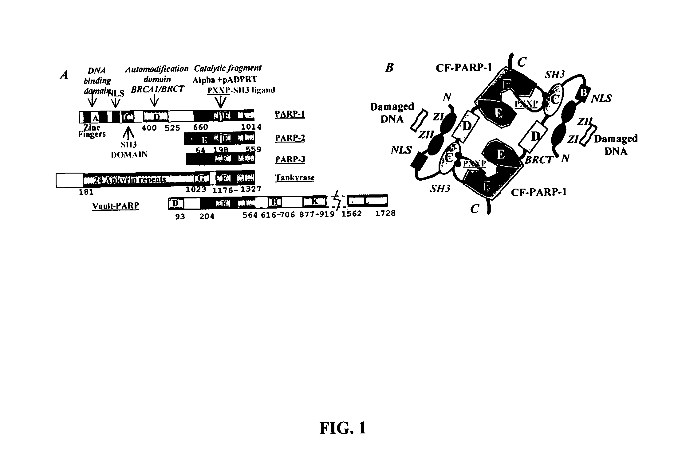
[0076]The binding of PARP-1 to DNA strand breaks results in its activation by the interaction of SH3 and SH3-ligand domains. This model provides the rationale for the development of a new drug discovery paradigm for generation of PARP-1 inhibitors for cancer therapy.
[0077]Functional Test of the Model Regarding the SH3 and SH3-ligand Domains of PARP-1. Directed mutagenesis is used to generate single amino acid changes to disrupt the peptide recognition surface of the PARP-1 SH3 domain and the SH3-ligand domain. Disruption of either domain results in a PARP-1 that is unable to be activated by DNA strand breaks.
[0078]Human PARP-1 SH3 is mutated at residues that map to a conserved, and predominately aromatic surface (L293A, P294A, C295A, W318A, W333A, P336A and F339A) involved in the recognition of the proline-rich PXXP SH3-ligand (PPII). The site-directed mutants are designed to maintain structural integrity of the SH3 domain, while disrupting its ability to i...
example 3
Molecular Modeling in Designing PARP-1 Inhibitors
[0091]Based upon the now determined structure of the SH3 domain and SH3-ligand domain of PARP-1, known and predicted compounds can be tested by molecular simulations for interaction with PARP-1, using InsightII (Molecular Visualization (MolViz) Facility Department of Chemistry Indiana University, Bloomington, Ind. USA) and other modeling software.
[0092]First, the structure of the PARP-1 SH-3domain or the PARP-1 SH3-ligand domain in a digital format that can be used by a molecular modeling computer program. The molecular modeling software generally provides information regarding which digital format is acceptable for that program. Next, the structure of a compound suspected of molecularly interacting with the PARP-1 SH-3domain or the PARP-1 SH3-ligand domain is obtained. The “molecularly interacting” can be covalent, ionic or other noncovalent binding. Guidance for how the compound may molecularly interact with these domains is provide...
PUM
| Property | Measurement | Unit |
|---|---|---|
| affinity | aaaaa | aaaaa |
| electrophoretic mobility | aaaaa | aaaaa |
| cellular resistance | aaaaa | aaaaa |
Abstract
Description
Claims
Application Information
 - R&D
- Intellectual Property
- Life Sciences
- Materials
- Tech Scout
- Unparalleled Data Quality
- Higher Quality Content
- 60% Fewer Hallucinations
Browse by: Latest US Patents, China's latest patents, Technical Efficacy Thesaurus, Application Domain, Technology Topic, Popular Technical Reports.
© 2025 PatSnap. All rights reserved.Legal|Privacy policy|Modern Slavery Act Transparency Statement|Sitemap|About US| Contact US: help@patsnap.com


