Preparation method of novel tissue engineering skin
A tissue-engineered skin, a new type of technology, applied in biochemical equipment and methods, tissue culture, epidermal cells/skin cells, etc., can solve the problem of immunogenicity of natural scaffolds that do not have biological activity, and achieve the effect of low immunogenicity
- Summary
- Abstract
- Description
- Claims
- Application Information
AI Technical Summary
Problems solved by technology
Method used
Image
Examples
Embodiment 1
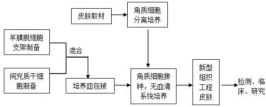
[0026] Embodiment 1 The present invention is realized through the following steps.
[0027] 1. Skin tissue preparation:
[0028] (1) Quickly place the excised autologous skin in sterile PBS solution and transport it to the laboratory at 4°C;
[0029] (2) Use scissors to remove the subcutaneous fat tissue as much as possible;
[0030] (3) Disinfect the skin tissue treated in step (2) with iodophor for 10 minutes at room temperature;
[0031] (4) Wash the sterilized skin tissue 3 times with sterile PBS;
[0032] (5) Place the skin tissue cleaned in step (4) in a sterile petri dish, and cut into small pieces with a size of 5mm×5mm;
[0033] (6) Put the skin tissue from step (5) in 15ml of discrete enzyme, and store it overnight at 4°C.
[0034] 2. Preparation of amniotic membrane decellularized scaffold:
[0035] (1) Disinfection: After separating the amniotic membrane, put it into a centrifuge tube, add 20ml of alcohol with a volume fraction of 65-75% per 15g of amniotic me...
Embodiment 2
[0052] Embodiment 2 The present invention is realized through the following steps.
[0053] 1. Skin tissue preparation:
[0054] (1) Quickly place the excised autologous skin in sterile PBS solution and transport it to the laboratory at 4°C;
[0055] (2) Use scissors to remove the subcutaneous fat tissue as much as possible;
[0056] (3) Disinfect the skin tissue treated in step (2) with iodophor for 10 minutes at room temperature;
[0057] (4) Wash the sterilized skin tissue 3 times with sterile PBS;
[0058] (5) Place the skin tissue cleaned in step (4) in a sterile petri dish, and cut into small pieces with a size of 5mm×5mm;
[0059] (6) Put the skin tissue from step (5) in 15ml of discrete enzyme, and store it overnight at 4°C.
[0060] 2. Preparation of amniotic membrane decellularized scaffold:
[0061] (1) Disinfection: After separating the amniotic membrane, put it into a centrifuge tube, add 20ml of alcohol with a volume fraction of 65-75% per 15g of amniotic me...
Embodiment 3
[0078] Embodiment 3 The present invention is realized through the following steps.
[0079] 1. Skin tissue preparation:
[0080] (1) Quickly place the excised autologous skin in sterile PBS solution and transport it to the laboratory at 4°C;
[0081] (2) Use scissors to remove the subcutaneous fat tissue as much as possible;
[0082] (3) Disinfect the skin tissue treated in step (2) with iodophor for 10 minutes at room temperature;
[0083] (4) Wash the sterilized skin tissue 3 times with sterile PBS;
[0084] (5) Place the skin tissue cleaned in step (4) in a sterile petri dish, and cut into small pieces with a size of 5mm×5mm;
[0085] (6) Put the skin tissue from step (5) in 15ml of discrete enzyme, and store it overnight at 4°C.
[0086] 2. Preparation of amniotic membrane decellularized scaffold:
[0087] (1) Disinfection: After separating the amniotic membrane, add it to a centrifuge tube, add 20ml of alcohol with a volume fraction of 65-75% for each 15g of amniotic...
PUM
 Abstract
Description
Claims
Application Information
 - R&D
- Intellectual Property
- Life Sciences
- Materials
- Tech Scout
- Unparalleled Data Quality
- Higher Quality Content
- 60% Fewer Hallucinations
Browse by: Latest US Patents, China's latest patents, Technical Efficacy Thesaurus, Application Domain, Technology Topic, Popular Technical Reports.
© 2025 PatSnap. All rights reserved.Legal|Privacy policy|Modern Slavery Act Transparency Statement|Sitemap|About US| Contact US: help@patsnap.com


