
Implantable medical device employing sonomicrometer output signals for detection and measurement of cardiac mechanical function

a sonomicrometer and output signal technology, applied in the field of implantable medical devices, can solve the problems of insufficient cardiac output to sustain mild or moderate levels, inability to distinguish a true sense event in a distorted signal, and insufficient cardiac outpu

Inactive Publication Date: 2006-07-25
MEDTRONIC INC
View PDF56 Cites 50 Cited by
  • Summary
  • Abstract
  • Description
  • Claims
  • Application Information

AI Technical Summary

Benefits of technology

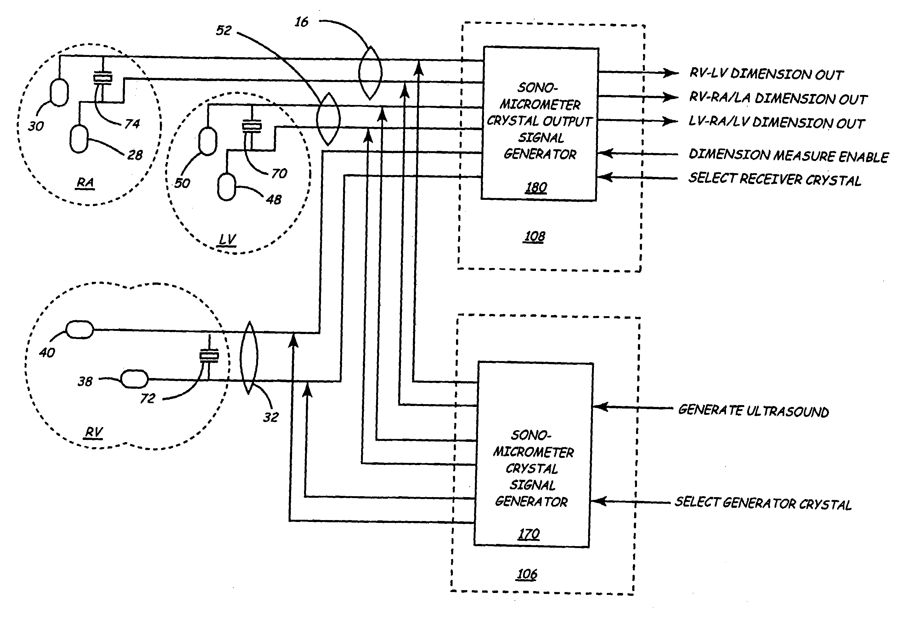
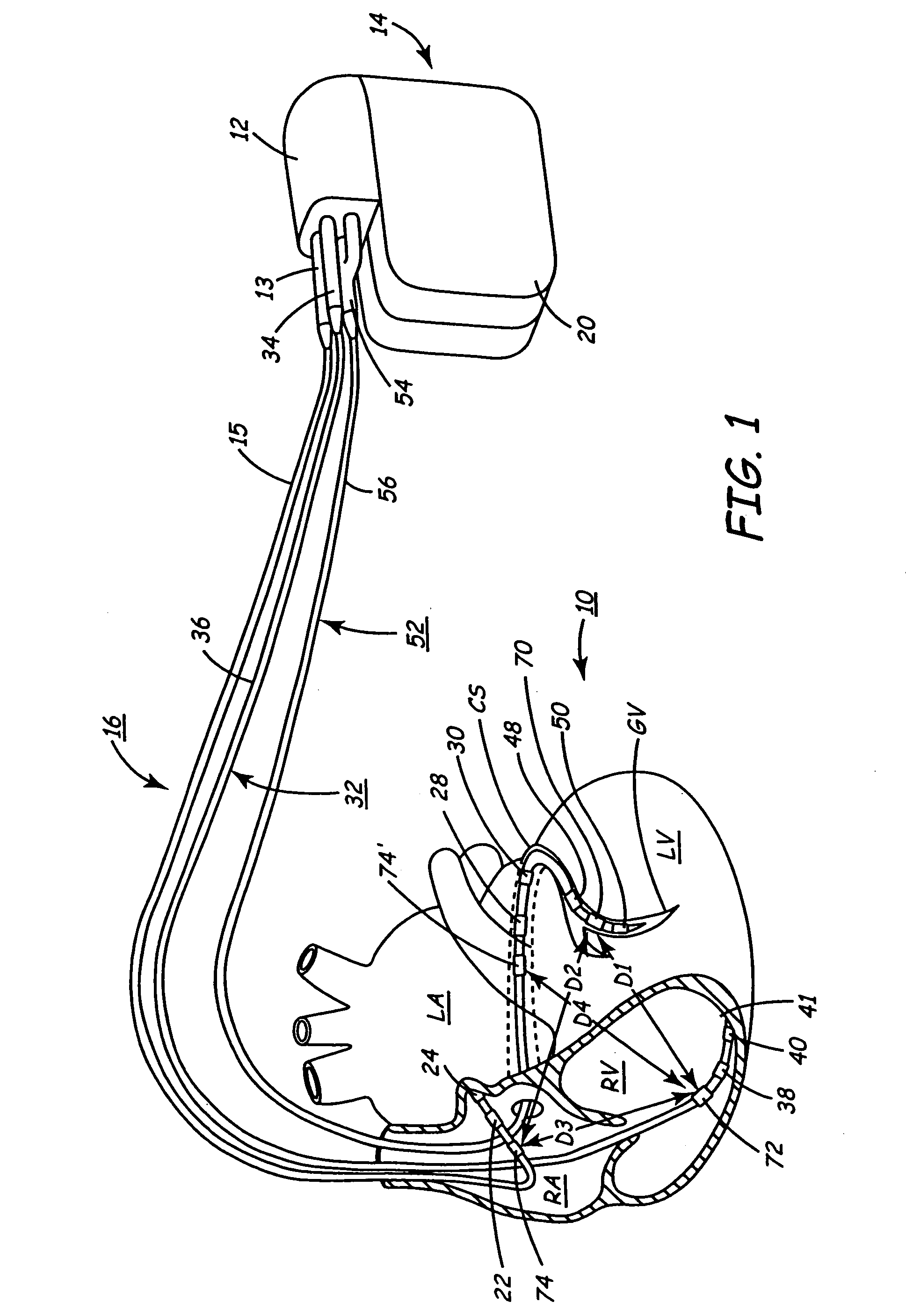
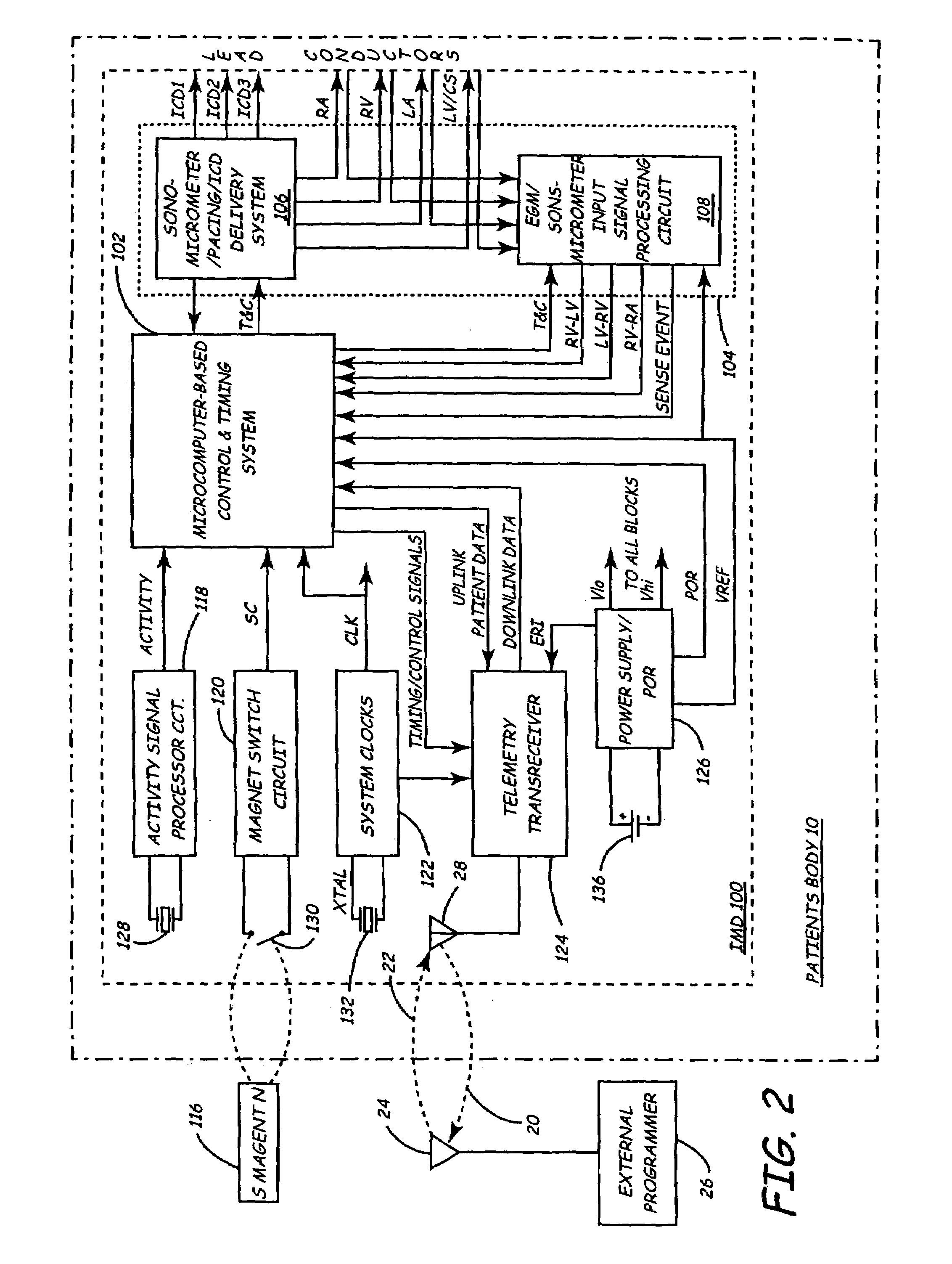
[0018]In view of the above need, the present invention provides a system and method incorporated into therapy delivery and / or monitor IMDs employing dimension sensors for determining mechanical heart function and measuring mechanical heart performance of both left and right heart chambers without intruding into a left heart chamber or requiring invasive surgery to access the left heat chamber epicardium.
[0023]A principal advantage of the system and method of the present invention include the ability to directly monitor left ventricular function without entering the left side of the heart, the ability to monitoring atrial mechanical function, and the potential for parallel wiring with pace / sense electrodes, thereby avoiding the need for additional conductors within the lead body and connector elements.

Problems solved by technology

It was proposed that blood pressure sensors or accelerometers be incorporated so that the absence of mechanical heart function during fibrillation could also be detected to confirm the tentative determination of fibrillation before a shock therapy was delivered, but suitable sensors were not available.
The ability to distinguish a true sense event in a distorted signal is also complicated by the necessity of protecting sense amplifier circuitry following delivery of a pacing pulse or cardioversion / defibrillation shock.
These disease processes lead to insufficient cardiac output to sustain mild or moderate levels of exercise and proper function of other body organs, and progressive worsening eventually results in cardiogenic shock, arrhythmias, electromechanical dissociation, and death.
However, the measurement procedure is time consuming to perform for even a resting patient and cannot be practically performed replicating a range of metabolic conditions.
Typically, the echocardiography procedure is performed infrequently and months or years may lapse between successive tests, resulting in a poor understanding of the progress of the disease or whether or not intervening drug therapies have been efficacious.
However, it is asserted that the two-crystal system has several disadvantages such as (1) it is necessary to insert a piezoelectric crystal through the myocardium, which can damage the myocardial nerve and blood vessel supply of the myocardial wall, (2) it is difficult to position precisely the endocardial crystal at the tissue / blood sub-endocardial interface, and (3) it can be difficult to maintain good alignment at all times throughout the cardiac cycle for short term and particularly during longer duration studies (>12 weeks).
The introduction of such sensors into left heart chambers through the arterial system introduces complications that may be difficult to manage both acutely and chronically.
The surgical approach to the exterior of the heart is also not favored as it complicates the surgery and recovery of the patient.
The prior mechanical heart function sensors devices have been generally unsuccessful due to a.) the added complexity and reduced reliability of associated leads, and b.) the challenge of obtaining useful hemodynamic information from such surrogate measurements, which usually address only the right heart chambers.

Method used


Image

  • Implantable medical device employing sonomicrometer output signals for detection and measurement of cardiac mechanical function
  • Implantable medical device employing sonomicrometer output signals for detection and measurement of cardiac mechanical function
  • Implantable medical device employing sonomicrometer output signals for detection and measurement of cardiac mechanical function

Examples


Embodiment Construction

[0047]In the following detailed description, references are made to illustrative embodiments for carrying out the invention. It is understood that other embodiments may be utilized without departing from the scope of the invention. For example, the invention is disclosed in detail herein in the context of an AV sequential, three chamber or four chamber, pacing system operating in demand, atrial tracking, and triggered pacing modes for restoring synchrony in depolarizations and contraction of left and right ventricles in synchronization with atrial sensed and paced events for treating heart failure and / or bradycardia in those chambers. This embodiment of the invention is programmable to operate as a three or four chamber pacing system having an AV synchronous operating mode for restoring upper and lower heart chamber synchronization and right and left atrial and / or ventricular chamber depolarization synchrony.

[0048]It should be appreciated that the present invention may be utilized i...


PUM


Abstract

Implantable medical devices (IMDs) for detection and measurement of cardiac mechanical and electrical function employ a system and method for determining mechanical heart function and measuring mechanical heart performance of upper and lower and left and right heart chambers without intruding into a left heart chamber through use of a dimension sensor. The dimension sensor or sensors comprise at least a first sonomicrometer piezoelectric crystal mounted to a first lead body implanted into or in relation to one heart chamber that operates as an ultrasound transmitter when a drive signal is applied to it and at least one second sonomicrometer crystal mounted to a second lead body implanted into or in relation to a second heart chamber that operates as an ultrasound receiver.

Description

RELATED APPLICATION[0001]This application is a continuation of U.S. Pat. No. 10 / 002,338 filed Oct. 30, 2001 now U.S. Pat. No. 6,795,732, entitled “IMPLANTABLE MEDICAL DEVICE EMPLOYING SONOMICROMETER OUTPUT SIGNALS FOR DETECTION AND MEASUREMENT OF CARDIAC MECHANICAL FUNCTION”, to Stadler et al., incorporated herein by reference in its entirety.FIELD OF THE INVENTION[0002]The present invention relates generally to implantable medical devices (IMDs) that deliver therapies to the heart and / or monitor cardiac physiologic parameters that, in particular, involves implantation of sonomicrometer piezoelectric crystals in or in relation to the heart chambers to detect occurrence of and magnitudes of the mechanical movements of the heart chambers.BACKGROUND OF THE INVENTION[0003]A wide variety of IMDs have been developed over the years or are proposed that provide cardiac rhythm management of disease states manifested by cardiac rhythm disorders and heart failure. Implantable pacemakers have b...

Claims


Application Information

Patent Timeline
IPC IPC(8): A61N1/365A61B8/00A61N1/362
CPCA61B8/00A61N1/36528A61N1/3627A61N1/36578
Inventor STADLER, ROBERT W.COMBS, WILLIAM J.LIPSON, DAVID
Owner MEDTRONIC INC
Learn More