Serum-free medium for mesenchymal stem cells

a mesenchymal stem cell and serum-free technology, applied in the field of serum-free medium for mesenchymal stem cells, can solve the problems of high variability between animal serum batches, time-consuming and labor-intensive preparation of autologous serum from patients, and certain disadvantages and limitations

Inactive Publication Date: 2006-09-19
CONSORZIO PER LA GESTIONE DEL CENT DI BIOTECNOLOGIA AVANZATA +1
View PDF6 Cites 67 Cited by
  • Summary
  • Abstract
  • Description
  • Claims
  • Application Information

AI Technical Summary

Problems solved by technology

However, the presence of animal or autologous serum in chondrocyte and MSC cultures has certain disadvantages and limitations in view of the potential therapeutical applications of these cultures.
Further, there is often high variability between animal serum batches.
Extensive serum screening required to select the batch most representative of the in vivo inductive effects can be time-consuming and expensive The preparation of autologous serum from patients is also time consuming and supplies are limited.
Animal serum can further potentially carry unknown pathogens with consequent risk of contamination for the patient.

Method used


Image

  • Serum-free medium for mesenchymal stem cells

Examples


example 1

[0028]Articular cartilage was harvested from the knee joint of young adult human donors. The samples were first cleaned of any adherent muscular, connective or subchondral bone tissues, minced into 1–3 mm fragments and rinsed in PBS. Single chondrocytes were then released by repeated enzymatic digestions at 37° C. with 0.25% trypsin, 400 U / ml collagenase I, 1000 U / ml collagenase II and I mg / ml hyaluronidase. Trypsin was then blocked and removed by rapid and extensive washes in PBS containing soybean trypsin inhibitor. Cells were plated in anchorage-dependent conditions in Coon's modified Ham's F-12 medium supplemented either with 10% fetal calf serum (FCS, control culture) or the following defined components: EGF, PDGFbb and FGF-2, ascorbic acid, linoleic acid, human serum albumin (HSA), β-mercaptoethanol, dexamethasone, insulin, human holo- and apo-transferrin. Table 1 below shows the preferred amounts of each component. To favor adhesion of the cells in serum free conditions, the ...

example 2

[0031]Bone marrow sample harvested from the iliac crest of the patient was washed twice with PBS. The nucleated cells were counted using methyl violet and plated at 5×10⁶ cells as unfractionated marrow per 10 cm tissue culture dish. For selection and expansion, the cells were maintained in Coon's modified Ham's F12 (F12) supplemented either with 10% FCS and 1 ng / ml FGF-2 (control culture) or the following defined components: EGF, PDGFbb, FGF-2, LIF, SCF, IGF-I, ascorbic acid, cholesterol, HSA, β-mercaptoethanol, dexamethasone, human holo- and apo-transferrin, selenium, biotin and sodium pantotenate. Table 2 below shows the preferred amounts of each component. To favor adhesion of the MSCs, the cells were first plated for 48 hours in F12 medium supplemented with 10% human serum and 1 ng / ml FGF-2. Thereafter, the medium was removed and the cells were extensively washed with PBS, and left for an additional 24–48 hours in F12 medium without any supplement. The defined mixture of factors...

example 3

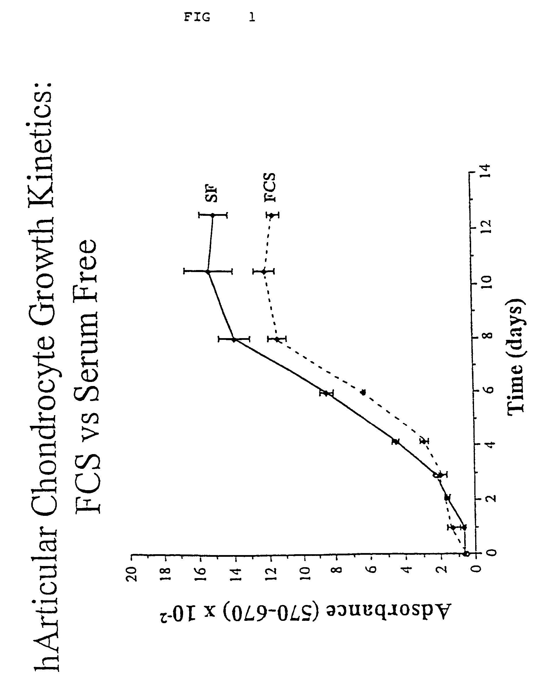
[0035]Growth Kinetics of Chondrocytes

[0036]At day 0, 5×10³ first passage cells were plated in each well of a 24-well plate in the presence of FCS. Upon adhesion, the FCS was removed, and the cells were extensively washed with PBS and left for 2–3 days in F12 without supplement to exhaust residual traces of serum. Proliferation was then reinduced by adding either 10% FCS or the mixture of defined components established for chondrocytes. Cell number was evaluated at different days via Thiazolyl blue (MTT) staining. Briefly, culture medium was removed and replaced with 0.5 ml of medium without supplement; then 25 μl MTT (Sigma, St. Louis, Mo.) stock solution (5 mg / ml) was added to each culture being assayed. After a 3 hour incubation the medium was removed and the converted dye solubilized with absolute ethanol. Absorbance of converted dye was measured at a wavelength of 570 nm with background subtraction at 670 nm.

[0037]The data obtained (see FIG. 1) clearly show that the defined medi...


PUM

PropertyMeasurementUnit
concentrationsaaaaaaaaaa
concentrationaaaaaaaaaa
concentrationsaaaaaaaaaa

Abstract

Serum-free media for growth and proliferation of chondrocytes and mesenchymal stem cells in culture are provided. A serum-free medium for growth of chondrocytes includes a serum-free composition comprising FGF-2, linoleic acid, ascorbic acid, B-mercaptoethanol, transferrin and dexamethasone. Further, the composition comprises EGF, PDGFbb, insulin and albumin. A method for growing chondrocytes in a serum free medium comprising the compostion is also provided. Also provided for mesenchymal stem cell growth, is a serum-free medium which includes a composition comprising FGF-2, LIF, SCF, pantotenate, biotin and selenium and method, therefore.

Description

[0001]The present application is divisional of application Ser. No. 09 / 831,161, filed Jun. 11, 2001 now U.S. Pat. No. 6,617,159 which is a 371 U.S. National Phase of PCT / EP99 / 08482, filed 8 Nov. 1999, which claims benefit of U.S. Provisional application Ser. No. 60 / 107,646, filed 9 Nov. 1998, the entire contents of each of which is incorporated herein by reference.BACKGROUND OF THE INVENTION[0002]Bone and cartilage transplantation is an absolute need in reconstruction of bone and cartilage segments in plastic surgery, traumatic surgery or after the removal of neoplastic lesions, etc. Typically, material of human (autologous, from donors or from cadavers) or animal origin has been used for this purpose. Given the increased demand from clinicians for transplant tissues, the increased need for microbial safety in tissue transplantation, the advances in cell biology, cell differentiation and tissue engineering, the concept of rebuilding tissues from autologous or allogeneic cells expand...

Claims


Application Information

Patent Timeline
Patent Type & Authority Patents(United States)
IPC IPC(8): C12N5/00C12N5/02A61K35/12C12N5/077C12N5/0775C12R1/91
CPCC12N5/0655C12N5/0663A61K35/12C12N2500/25C12N2500/36C12N2500/38C12N2500/99C12N2501/11C12N2501/115C12N2501/125C12N2501/135C12N2501/235C12N2501/39C12N2501/105C12N2500/90
Inventor CANCEDDA, RANIERIDOZIN, BEATRICE
Owner CONSORZIO PER LA GESTIONE DEL CENT DI BIOTECNOLOGIA AVANZATA
Why Eureka
Try Eureka