Nucleic acid sensor molecules and methods of using same

a sensor molecule and nucleic acid technology, applied in the field of nucleic acids, can solve the problems of antibody-based detection methods, unstable monoclonal antibody fragments, and limited utility of antibody methods such as elisa or competitive ria, and achieve the effect of changing the optical properties of said molecule, increasing the detectable fluorescence of said fluorescent donors

Inactive Publication Date: 2006-10-24
ARCHEMIX CORP
View PDF116 Cites 86 Cited by
  • Summary
  • Abstract
  • Description
  • Claims
  • Application Information

AI Technical Summary

Benefits of technology

[0025]In one embodiment, the nucleic acid sensor molecule of the invention has an optical signal generating unit that includes at least one signaling moiety. In another embodiment, the nucleic acid sensor molecule of the invention has an optical signal generating unit which includes at least a first signaling moiety and a second signaling moiety. In another embodiment, the first and second signaling moieties change proximity to each other upon recognition of a target by the target modulation domain. In another embodiment, the first and second signaling moieties include a fluorescent donor and a fluorescent quencher, and recognition of a target by the target modulation domain results in an increase in detectable fluorescence of said fluorescent donor. In another embodiment, the first signaling moiety and said second signaling moiety include fluorescent energy transfer (FRET) donor and acceptor groups, and recognition of a target by the target modulation domain results in a change in distance between said donor and acceptor groups, thereby changing optical properties of said molecule.

Problems solved by technology

A limitation with this type of aptamer-derived biosensor is that ligand-mediated changes in secondary structure were engineered into the aptamer molecule via a laborious engineering process in which four to six nucleotides were added to the 5′ end of the aptamer that was complementary to the bases at the 3′ end of the thrombin binding region.
In addition, antibody methods such as ELISA or competitive RIA, while robust, are restricted in utility because these methods require that heterogeneous assay conditions be employed: 1] detection is done on a solid surface; 2] in most applications both a capture antibody and detection antibody are required; 3] for ELISA-based protein detection methods, the antibodies must recognize the folded, native structure of the protein that is present in cell or tissue isolates and; 4] antibody and protein based detection methods have not been described for intracellular or in vivo based analyte detection.
First monoclonal antibody fragments are unstable and do not fold properly when expressed as intracellular protein molecules.
Second, intracellular detection requires homogeneous assay formats and these solution-based detection methods require the sensor to have a ligand sensing or modulation domain coupled directly to a catalytic or signal generating region of an enzyme or catalytic biomolecule.
A fundamental and important consequence of the limitations of antibody-based detection methods is that they can not function as a universal reagent for all assays and tests that can be employed in drug discovery and development.
Another limitation of the engineering method to sensor generation is that it has been generally thought not to be applicable to the development of protein-dependent ribozymes.
Furthermore, the authors state that the “principles required for engineering protein-dependent ribozymes must be fundamentally different from those for identifying ribozymes dependent on small-molecules.” Hence, in order to identify protein and peptide dependent ribozymes, Ellington and Robertson undertook a laborious in vitro selection process which involved randomization of the catalytic core of the L1 ligase coupled with multiple rounds of positive and negative selection.
First, the nucleoprotein enzymes developed by Ellington and Robertson (2000) required a laborious in vitro selection process to identify peptide and protein dependent ligases.
Hence, a hindrance to the development of L1 ligase-based biosensor technology is the lack of a general method for the generation of biosensors that can work in multiple assay and detection formats required of solution-based and chip-based biosensors and, those that can work in multiplexed formats and in complex biological extracts.

Method used


Image

  • Nucleic acid sensor molecules and methods of using same
  • Nucleic acid sensor molecules and methods of using same
  • Nucleic acid sensor molecules and methods of using same

Examples


example

Example 1

General Procedures

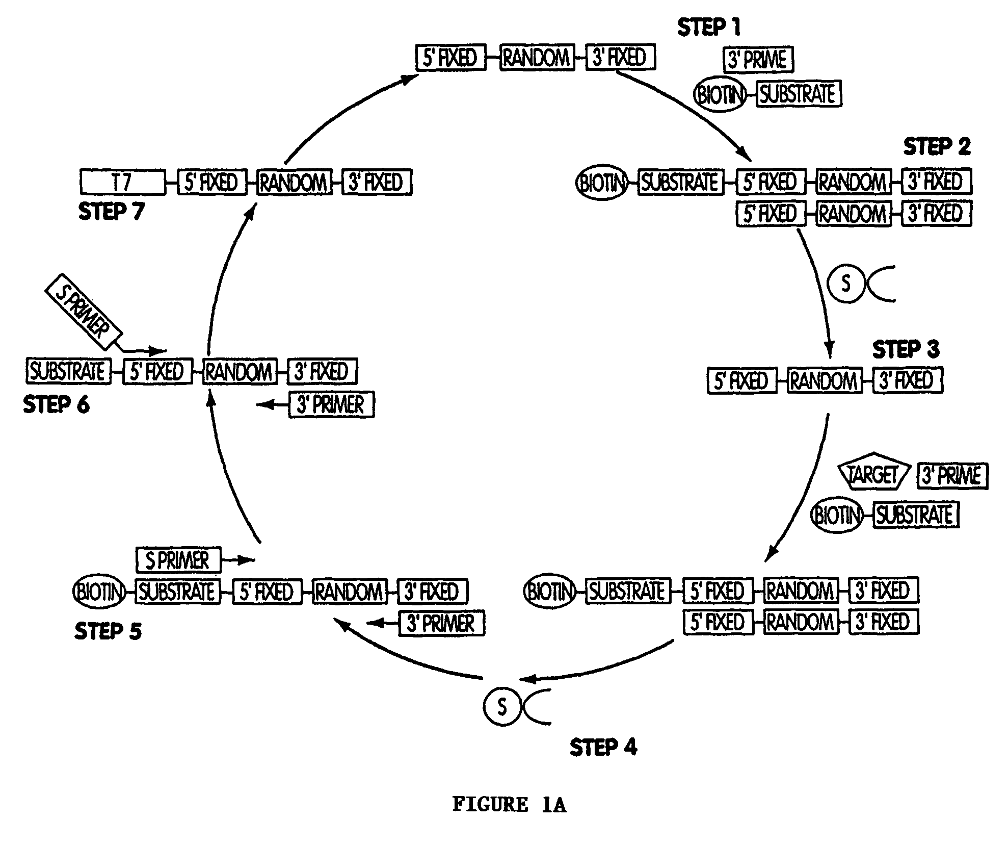
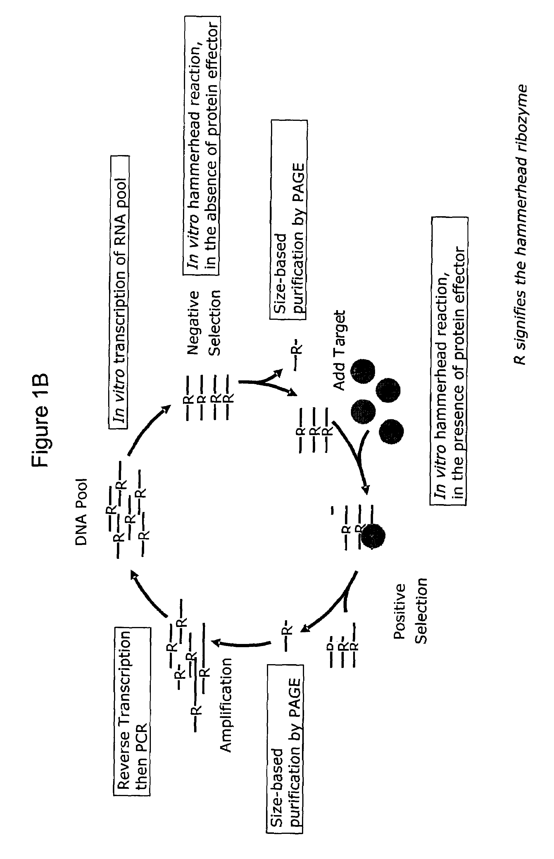
[0441]A. Generation nucleic acid sensor molecules from pools of ribozymes comprised of randomized linker and target modulation domains.

[0442]Direct selection of nucleic acid sensor molecules. Target modulated nucleic acid sensor molecules are isolated by in vitro selection. Pool of partially randomized ribozymes with 1015–1017 unique sequences serve as the starting point for in vitro selection. As with the engineering approach, both the L1 ligase and hammerhead ribozyme are used as platforms for the selection of allosterically-controlled molecules. Selections are designed to yield ribozymes that specifically respond to any target. Nucleic acid sensor molecules with cross-specificity (i.e. modulated by alternate target molecules, or by alternate ligand-bound states, or by alternate post-translationally modified forms of protein or peptides) are selected against by including the undesired form of the target in an initial negative selection step. The specific...

example 2

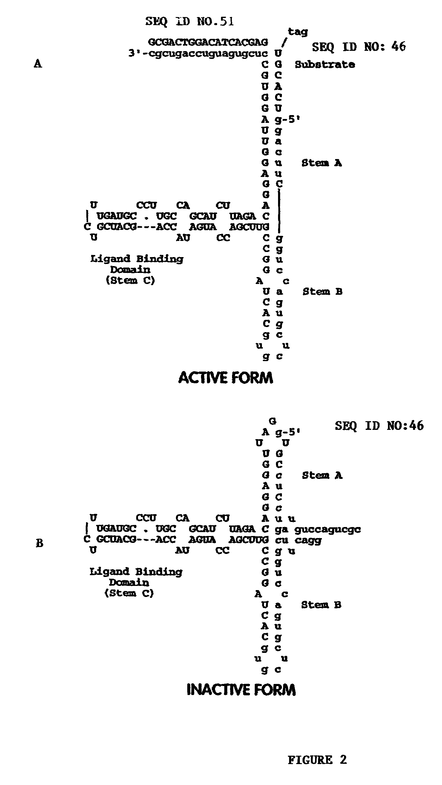
Selection for a Nucleic Acid Sensor Molecule Selective for the Estrogen Receptor LBD

[0483]A nucleic acid sensor molecule which is modulated by the estrogen receptor (ER) ligand binding domain (LBD) is obtained by in vitro selection methods to identify candidate nucleic acid sensor molecules that are modulated by an estrogen receptor LBD.

[0484]The full length gene for the estrogen receptor is known. One source of the full-length estrogen receptor clone is Acc. No. M12674 (see also Greene et al., Science 231:1150–54, 1986). The clone includes a 2092 nucleotide mRNA with the sequence presented in Table 3 below:

[0485]

TABLE 31gaattccaaa attgtgatgt ttcttgtatt tttgatgaag gagaaatact gtaatgatca(SEQ ID NO:9)61ctgtttacac tatgtacact ttaggccagc cctttgtagc gttatacaaa ctgaaagcac121accggacccg caggctcccg gggcagggcc ggggccagag ctcgcgtgtc ggcgggacat181gcgctgcgtc gcctctaacc tcgggctgtg ctctttttcc aggtggcccg ccggtttctg241agccttctgc cctgcgggga cacggtctgc accctgcccg cggccacgga ccatgaccat301gaccctccac accaa...

example 3

Selection for a Library of Nucleic Acid Sensor Molecules which Signal the Presence of all Known Nuclear Hormone Receptor LBDs:

[0492]A wide variety of nuclear hormone receptor (“NHR”) ligand binding domains and their ligands, many of which are described in Table 5 below, are known for which a nucleic acid sensor molecule can be selected.

[0493]

TABLE 5SymbolDescriptionLigandAIB3nuclear receptor coactivator RAP250; peroxisomethyroid hormoneproliferator-activated receptor interacting protein;thyroid hormone receptor binding proteinARandrogen receptor (dihydrotestosterone receptor;dihydroxytestosterontesticular feminization; spinal and bulbar muscularatrophy; Kennedy disease)C1Dnuclear DNA-binding proteinESR1estrogen receptor 1estrogenESR2estrogen receptor 2 (ER beta)estrogenESRRAestrogen-related receptor alphaestrogen and TFIIBESRRBestrogen-related receptor betaestrogen and TFIIBESRRGestrogen-related receptor gammaestrogen and TFIIBHNF4Ahepatocyte nuclear factor 4, alphaHNF4Ghepatocyte n...


PUM

PropertyMeasurementUnit
optical signalaaaaaaaaaa
detectable fluorescenceaaaaaaaaaa
fluorescent energy transferaaaaaaaaaa

Abstract

Methods for engineering a nucleic acid sensor molecule are provided. Biosensors comprise a plurality of nucleic acid sensor molecules labeled with a first signaling moiety and a second signaling moiety. The nucleic acid sensor molecules recognizes target molecules which do not naturally bind to DNA. Binding of a target molecule to the sensor molecules triggers a change in the proximity of the signaling moieties which leads to a change in the optical properties of the nucleic acid sensor molecules on the biosensor. Reagents and systems for performing the method are also provided. The method is useful in diagnostic applications and drug optimization.

Description

RELATED APPLICATIONS[0001]This application claims priority to U.S. Ser. No. 60 / 311,378, filed Aug. 9, 2001; U.S. Ser. No. 60 / 313,932, filed Aug. 21, 2001; U.S. Ser. No. 60 / 338,186, filed Nov. 13, 2001; U.S. Ser. No. 60 / 349,959, filed Jan. 18, 2002; U.S. Ser. No. 60 / 364,486, filed Mar. 13, 2002; U.S. Ser. No. 60 / 367,991, filed Mar. 25, 2002; U.S. Ser. No. 60 / 369,887, filed Apr. 4, 2002; U.S. Ser. No. 60 / 376,744, filed May 1, 2002; U.S. Ser. No. 60 / 385,097, filed May 31, 2002; U.S. Ser. No. 60 / 391,719, filed Jun. 26, 2002; and continuation-in-part U.S. Ser. No. 09 / 952,680, filed Sep. 13, 2001 now abandoned, which claims priority to U.S. Ser. No. 60 / 232,454, filed Sep. 13, 2000. The contents of these applications are incorporated herein by reference in their entirety.FIELD OF THE INVENTION[0002]The invention relates generally to nucleic acids and more particularly to nucleic acid sensor molecules containing a catalytically active domain that is modulated upon recognition of a target by...

Claims


Application Information

Patent Timeline
Patent Type & Authority Patents(United States)
IPC IPC(8): C12Q1/00C07H19/00C12Q1/68C07H21/02C07H21/04C12Q1/48C40B40/06C40B60/14
CPCB82Y30/00C07H21/02C07H21/04C12Q1/485C12Q1/6816C12Q1/6825B01J2219/00385B01J2219/00479B01J2219/00608B01J2219/0061B01J2219/00612B01J2219/00626B01J2219/0063B01J2219/00637B01J2219/00653B01J2219/00662B01J2219/00691B01J2219/00722C40B40/06C40B60/14C12Q2565/101C12Q2525/179C12Q2521/337C12Q2521/501
Inventor STANTON, MARTINEPSTEIN, DAVIDHAMAGUCHI, NOBUKOKURZ, MARKUSKEEFE, TONYWILSON, CHARLESGRATE, DILARAMARSHALL, KRISTIN A.MCCAULEY, THOMAS G.KURZ, JEFFREY C.
Owner ARCHEMIX CORP
Why Eureka
Try Eureka