

Methods for making and delivering rho-antagonist tissue adhesive formulations to the injured mammalian central and peripheral nervous systems and uses thereof
a technology of rho-antagonist and tissue adhesive, applied in the field of mammalian nervous system repair, can solve the problems of permanent functional impairment, deficits associated with spinal cord injury, loss of axons that are damaged, etc., and achieve the effect of preventing gel and c3 from lysis and improving the delivery of c3 in collagen
- Summary
- Abstract
- Description
- Claims
- Application Information
AI Technical Summary
Benefits of technology
Problems solved by technology
Method used
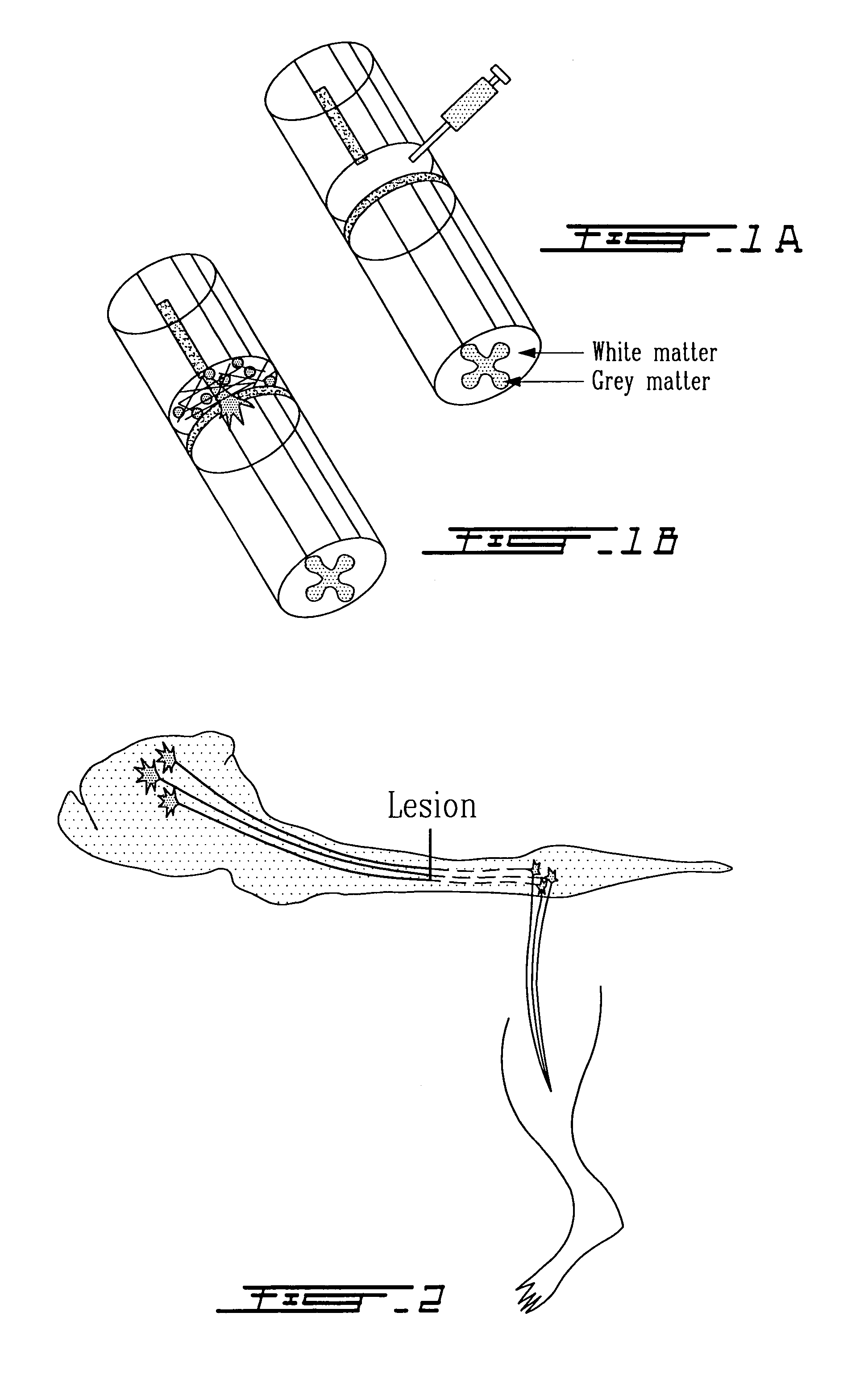
Image

Examples
example 1
A Kit for a Tissue Adhesive System
[0150]The kit contains:
1 vial aprotinin solution for reconstitution of fibrinogen
1 vial calcium chloride solution for reconstitution of thrombin
1 vial C3 solution
[0151]1.1 Lyophilized fibrinogen (75 mg / ml) in glycine buffer (2 mg / ml NaCl, 4 mg / ml trisodium citrate, 15 mg / ml glycine) was reconstituted in an aprotinin solution 3000 KIU / ml and heated to 37 C. For ease of handling, a combined heating and stirring device was used (appropriate vials contain a maganetic stirrer. This is called solution I.
[0152]1.2 A thrombin solution is prepared; the solution comprisin Lyophilized thrombin 500 IU / ml, 2.4 mg / ml glycine, 8 mg / ml sodium chloride. The calcium chloride solution (40 umol CaCl2) and thrombin are mixed and heated to 37C. This is called solution II.
[0153]1.3 A solution of C3 (1 mg / ml) is heated to 37C
[0154]1.4 Equal amounts of solution I, II, and III are mixed, and immediately drawn up in a syringe, and added to the ...
example 2
Modification of the Kit in Example 1
[0157]The formulation given in example 1 was used with the following modifications. Solution II is made with the addition of recombinant C3 directly to the solution II vial. In other words, solution II contains thrombin, calcium chloride and C3. Solution I is loaded in one syringe, solution II is loaded in a second syringe. A syringe with a plunger that simultaneously loads both solutions is used. Thus the solutions are mixed as they enter a small chamber before the needle, and the polymerization occurs in situ in the injured region of the CNS where the solution is applied. The system describe here is the Duploject system from Baxter Phamaceuticals U.S.A.
example 3
Modification of the Kit in Example 1
[0158]As example 2, but the C3 solution is mixed in vial 1 with the fibrinogen. Vial one and vial II are heated and prepared as described in example 1, and injected into the injured CNS with the Duploject system.
PUM
| Property | Measurement | Unit |
|---|---|---|
| distance | aaaaa | aaaaa |
| concentration | aaaaa | aaaaa |
| concentrations | aaaaa | aaaaa |
Abstract
Description
Claims
Application Information