
Probes for identifying cancer-specific antigens

a technology of antigens and probes, applied in the field of probes for identifying cancer-specific antigens, can solve the problems of reducing the ten-year survival rate, affecting the generation of suitable and effective therapies, and remained the major stumbling block to the identification of cancer-specific molecules,

Inactive Publication Date: 2008-02-12
CANCERPROBE
View PDF8 Cites 0 Cited by
  • Summary
  • Abstract
  • Description
  • Claims
  • Application Information

AI Technical Summary

Benefits of technology

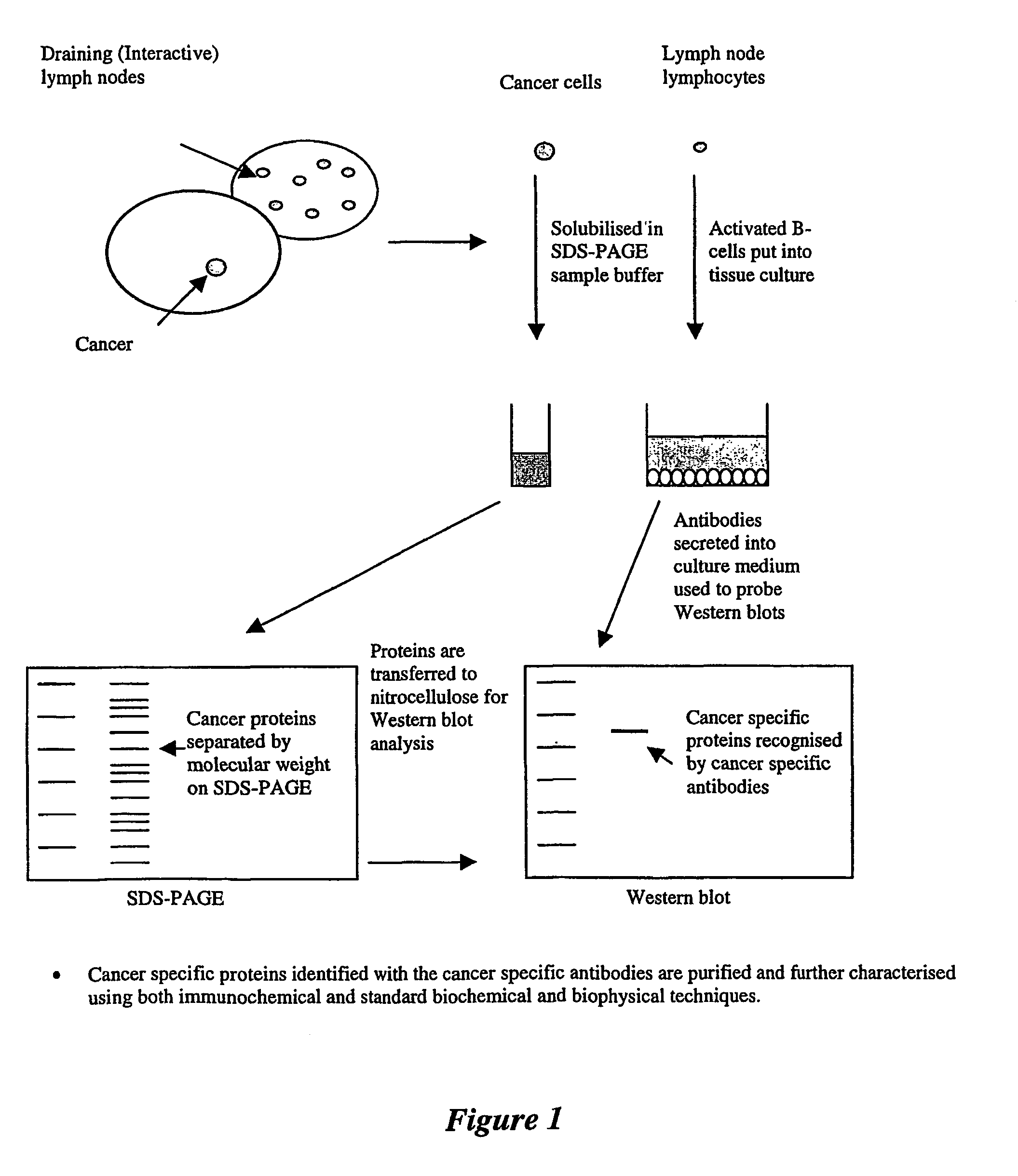
[0027]Because the level of antibody secretion into the culture medium may be very low without in vitro stimulation of resting lymphocytes, the method preferably includes a further step of activating the isolated cells to proliferate and to secrete antibodies, by adding a cell activating agent or a cell proliferation agent to the culture medium. In vitro secretion of antibodies into the culture medium by recently activated B cells may also be enhanced by the addition of mitogens or helper factors to the cultures.
[0042]The application of the ASC-probe of the invention to affinity chromatography enables sufficient quantities of cancer-specific antigens to be rapidly isolated from a complex crude extraction mixture for biochemical characterization, amino-acid sequencing and investigation of the ability of the antigens to elicit a protective immune response. The use of affinity chromatography for obtaining antigen(s) avoids the difficulties often encountered when applying conventional biochemical techniques to the purification of an antigen about which little or no data is known. It also obviates the need to raise polyclonal or monoclonal antibodies for the purpose of analytical affinity chromatography. Large-scale preparation may, however, require the preparation of polyclonal or monoclonal antibodies.

Problems solved by technology

The identification of cancer-specific molecules has remained the major stumbling block to the generation of suitable and effective therapies and diagnostics.
The search for these subtle variations is often driven by hope or inspiration, and entails considerable difficulty, expense and time.
No such method of early diagnosis is available for lung cancers, which can only be detected by radiography, which is usually carried out after symptoms become apparent, resulting in a much lower ten-year survival rate (approximately 13%).
The usefulness of serological tests for cancer diagnosis has however been disappointing, partly because cancer-specific or cancer-associated antigens have not been well defined and characterised.
In addition, the use of serum as a source of specific antibodies has several inherent disadvantages, including:1. The presence of large amounts of serum antibodies not related to the pathological agent, resulting in false positive results and background reactions;2. The formation of antigen-antibody complexes in the serum which can disguise the presence of low titre antibodies / antigens;3. The inability to detect antibodies produced locally at restricted mucosal sites, such as breast, colon and lung tissues, in the serum because of the massive dilution of these antibodies upon movement into the blood; and4. The likelihood of persistence of specific antibodies in the serum, even in the absence of current disease makes it difficult to differentiate between previous and current disease.
However, such methods are directed to making monoclonal antibodies against an unidentified antigen of a known cancer, rather than to identifying a cancer-specific antigen.
In addition, monoclonal antibody production requires complex in vitro fusion and selection proceedings; the monoclonal antibodies thus produced do not reflect the total polyclonal immune response mounted against the cancer by the B cells in the draining lymph nodes.
While the use of ASC-probes has proven to be a major breakthrough in research relating to vaccines against infectious diseases(Meeusen and Maddox, 1999), this technique has not yet been applied to cancer research.
However, there is no evidence as to whether this represents a specific response to a cancer-associated antigen, and the nature of these antigens has not been identified.

Method used


Image

  • Probes for identifying cancer-specific antigens
  • Probes for identifying cancer-specific antigens
  • Probes for identifying cancer-specific antigens

Examples


example 1

Preparation of ASC-Culture Supernatants

[0104]Lymph nodes draining the breast were collected after surgery and were teased gently in Dulbecco-modified Eagle's medium (DME) containing 10% heat-inactivated fetal calf serum (FCS) and antibiotics (400U / ml penicillin and 0.1 mg / ml streptomycin). Cells were collected, washed three times and resuspended to a final concentration of 5×10⁶ cells / ml in complete culture medium (DME supplemented with 400U / ml penicillin, 0.1 mg / ml streptomycin, 10% FCS, 2 mM glutamine). Pokeweed mitogen (Sigma Aldrich) was added at a concentration of 2.5 μg / ml to increase antibody secretion. Culture flasks containing 10 ml cell suspensions or 24-well culture plates containing 2 ml cell suspension per well were incubated at 37° C. in an atmosphere of 5% CO2 in air, and cell supernatants harvested 5-6 days later. Culture supernatants containing antibodies secreted by antibody-secreting cells (ASC) present in the lymph node cell suspensions were subsequently stored a...

example 2

Purification and Labelling of Antibodies from Cell Supernatants

[0105]Antibodies secreted by the ASC in culture supernatant were purified by affinity chromatography using a Protein-G column (Pharmacia HiTrap Affinity Column, 1 ml). The column was pre-equilibrated with loading buffer (20 mM sodium phosphate, pH 7.0). The cell supernatants were applied to the Protein G column, and the unbound proteins removed using 10 column volumes of loading buffer. Bound proteins were eluted from the column using 3 column volumes of elution buffer (0.1 M glycine / hydrochloric acid, pH 2.7). The eluate was neutralised using 100 μl 5 M tris / hydrochloric acid, pH 8.0. The purified antibody solution was dialysed extensively against phosphate buffered saline (PBS) before being stored at 4° C.

[0106]Control antibodies were purified from the serum of a patient who was found not to have breast cancer. The method of purification was identical to that used for the ASC supernatants.

Biotinylation of Purified Anti...

example 3

Western Blotting

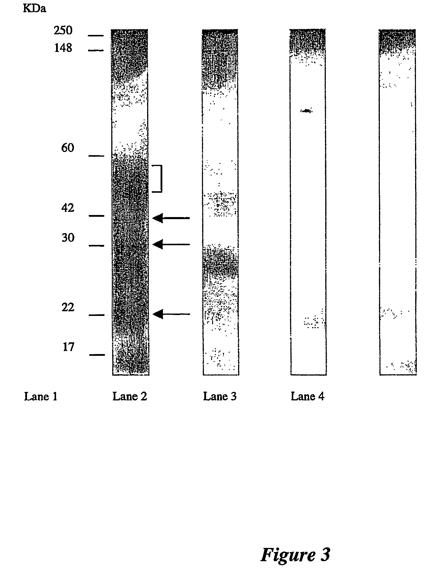
[0108]Appropriate dilutions of the antigens were loaded on a 10% SDS-PAGE gel and blotted on to nitrocellulose membrane (MSI, Melbourne, Australia). Western blot transfers were performed using the wet transfer apparatus (Bio-Rad Laboratories) at a constant voltage of 100V for 1 hour in transfer buffer (25 mM Tris-HCl pH 8.3, 192 mM glycine and 15% (v / v) methanol). After electrophoretic transfer, the membranes were placed in a blocking solution of 3% skim milk powder in PBS for 1 hour, then washed 3 times with 0.05% Tween 20 in PBS before being probed with antibody. For urine samples, Western blots were probed for 2 hours with undiluted ASC supernatant, followed by incubation for 1 hour with peroxidase-conjugated rabbit anti-human IgG serum (DAKO) at a 1:2000 dilution.

[0109]Western blots of breast tissue gave high reactions with the conjugate, due to contamination of the tissue with serum immunoglobulins. A one-step 2 hour incubation with purified biotinylated antibod...


PUM


Abstract

This invention relates to the diagnosis of cancer, and in particular to the identification and detection of cancer-specific antigens, the invention provides antibody probes and methods for using the probes for detection and purification of cancer-specific antigens, and in the preparation of vaccine compositions. Antibodies against cancer-specific antigens are also disclosed and claimed.

Description

[0001]This invention relates to the diagnosis of cancer, and in particular to the identification and detection of cancer-specific antigens. The invention provides antibody probes and methods for using the probes for detection and purification of cancer-specific antigens, and in the preparation of therapeutic, diagnostic and vaccine compositions, as well as in the preparation of antibodies directed against cancer-specific antigens.BACKGROUND OF THE INVENTION[0002]All references, including any patents or patent applications, cited in this specification are hereby incorporated by reference. No admission is made that any reference constitutes prior art. The discussion of the references states what their authors assert, and the applicants reserve the right to challenge the accuracy and pertinency of the cited documents. It will be clearly understood that, although a number of prior art publications are referred to herein, this reference does not constitute an admission that any of these ...

Claims


Application Information

Patent Timeline
IPC IPC(8): C12N5/06C12N5/08G01N33/48C07K16/30G01N33/574
CPCC07K16/3015C07K16/3069G01N33/574A61K2039/505
Inventor LOPATA, ALEXMEEUSEN, ELZAMANCUSO, NUNZIO
Owner CANCERPROBE
Learn More