Methods and compositions for nerve regeneration

a nerve regeneration and composition technology, applied in the field of methods and compositions for nerve regeneration, can solve the problems of limited treatment options for patients with spinal cord injuries, permanent impairment of neural function, damage to the spinal cord,

Inactive Publication Date: 2008-05-20
UNIVERSITY OF CHICAGO
View PDF7 Cites 4 Cited by
  • Summary
  • Abstract
  • Description
  • Claims
  • Application Information

AI Technical Summary

Benefits of technology

[0019]The inventor has further found that a different set of Wnt proteins pattern the connections of corticospinal tract (CST) axons projecting along the opposite direction by a repulsive mechanism. CST axons project from the motor cortex of the brain to the spinal cord motor circuits and send voluntary movement signals from the brain to the body. Several Wnt genes were found to be expressed at the dorsal funiculus in an anterior-to-posterior decreasing gradient at the cervical spinal cord, where CST axons first enter the spinal cord and a anterior-to-posterior increasing gradient at the lumbar spinal cord level, forming a “half-pipe” gradient. Wnt1 and Wnt5a can repel CST axons in collagen gel assays. A repulsive Wnt receptor, Ryk (Oshikawa et al., 2003; Halford et al., 2000), is expressed in the CST axons and can be detected at the pyramidal decussation and in the dorsal funiculus. Antibodies against the ectodomain of Ryk can block the repulsion of Wnt1. Finally, intrathecal injection of a Wnt inhibitor, secreted Frizzled related protein 2 (sFRP2), at the rostral cervical level (C1 and C2), can inhibit the posterior growth of CST axons in vivo, leading to weaker grip strength.
[0053]In some cases, it will be beneficial to apply one or more Wnt to the site of a spinal cord injury, such that the Wnt(s) will provide attractive guidance to those neurons that need to be attracted to the site of injury during regeneration and repellant guidance to those neurons that need to grow away from the site of injury during regeneration. In this regard, Wnt(s) applied at the site of an injury will provide directional guidance to axonal growth and cause sensory neurons to grow up through the site of the injury and repel motor neurons to grow down through the site of the injury. Further, in this embodiment, it may be beneficial to inhibit the Ryk pathway at the site of the injury so that motor neurons growing through the site of the injury are not inhibited by any Wnts present in the injury site, whether those Wnts are applied to the injury site, or expressed there as a result of normal adult Wnt expression or injury-induced Wnt expression. One may also apply a blocker of myelin inhibitors to the injury site, to prevent such inhibitors from impacting the growth of sensory neurons through the site.

Problems solved by technology

Unlike the peripheral nervous system, damage to the central nervous system axons, such as spinal cord axons cannot be repaired, causing permanent impairment of neural function, such as in paralysis.
The majority of broken necks and broken backs, or vertebral fractures, do not cause any spinal cord damage; however, in 10-14% of the cases where a vertebral trauma has occurred, the damage is of such severity it results in damage to the spinal cord.
Treatment options for patients with spinal cord injuries are limited.
Often, patients with SCI are left with severe, permanent disabilities.
A major reason for the limited availability of treatment options is the fact that there is little known about factors that can control and modulate nerve growth and regeneration following spinal cord injury.
The expression pattern of the Slits and Semaphorins identified in these studies have been examined, but no anterior-posterior gradient of these chemorepellents in the spinal cord has been identified, suggesting that these repellents do not control anterior-posterior pathfinding.
However, a role for Wnts in mammalian directional axonal growth regulation in the spinal cord has not previously been suggested or considered.

Method used


Image

  • Methods and compositions for nerve regeneration
  • Methods and compositions for nerve regeneration
  • Methods and compositions for nerve regeneration

Examples


example 1

Materials and Methods

[0219]Collagen gel assays. E13 rat spinal cord explants were cultured in collagen gel matrix as described previously (Tessier-Lavigne et al., 1988; Zou et al., 2000). These explants are either “open-book” or post-crossing or pre-crossing for the spinal cord commissural axons. COS7 cells were transfected with various expression constructs with FuGene6 reagent (Roche). The explants were typically cultured for 16-20 hours and fixed in 4% PFA for two hours. The “open-book” explants were analyzed by lipophilic DiI labelling using iontophoresis. The post-crossing explants were stained with a monoclonal antibody (E7) against β3 tubulin (Hybridoma Bank for Developmental Studies). The pre-crossing explants were stained with a monoclonal antibody (4D7) against TAG-1 (Hybridoma Bank for Developmental Studies). Both antibodies were detected using secondary antibodies conjugated with horseradish peroxidase and visualized with 3,3′-diaminobenzene (DAB) (Sigma). Quantification...

example 2

The A-P Guidance Cue(s) is Diffusible

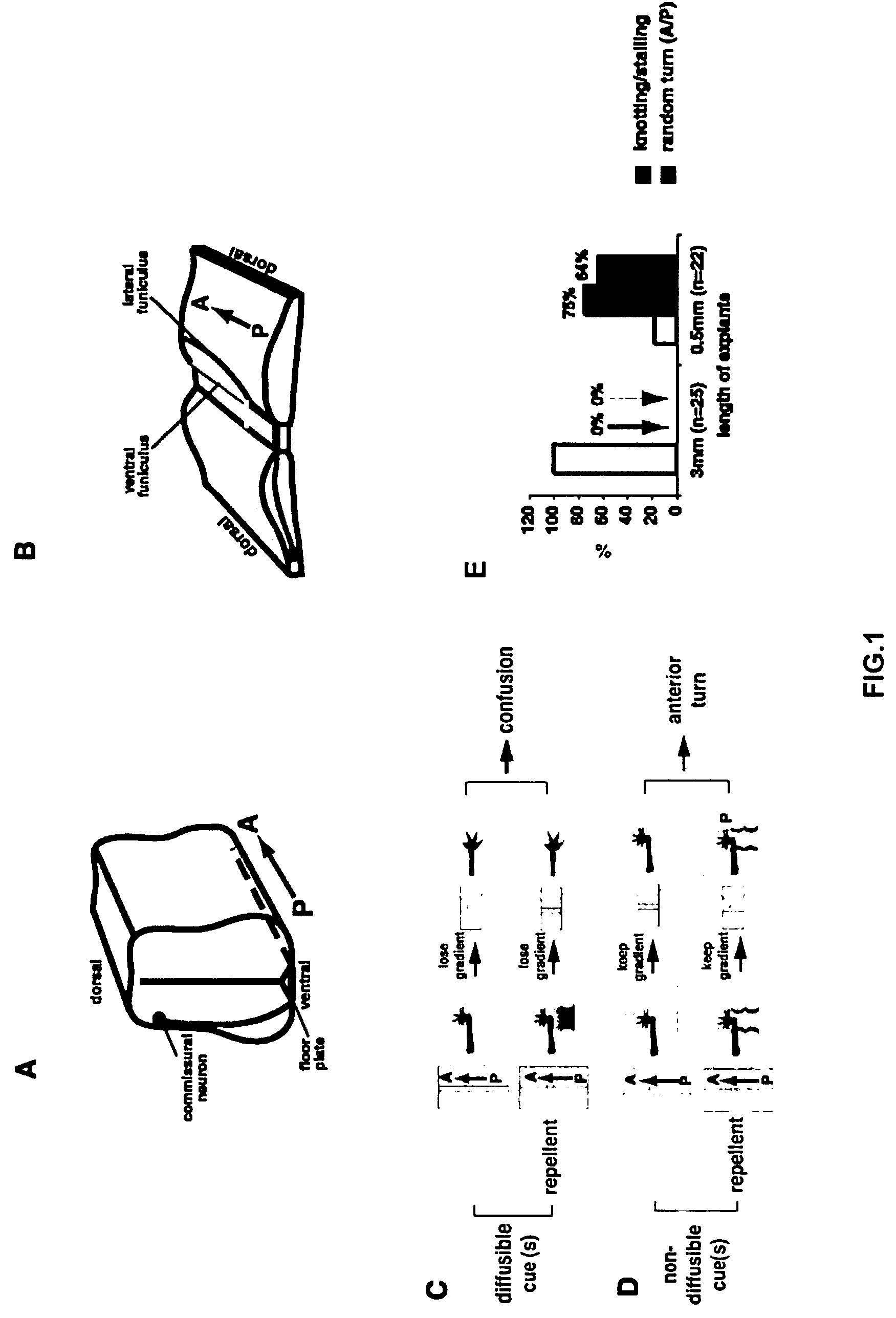
[0226]When a segment of E13 rat spinal cord is cultured in collagen gel for 16-18 hours, commissural axons were observed to project ventrally, cross the midline and turn anteriorly within the explant, mimicking their in vivo pathfinding. Commissural axon trajectories in these “open-book” explants can be revealed by lipophilic DiI injection into the dorsal side of the explants by iontophoresis (Fraser, 1996). Most of the commissural axons in E13 rat spinal cord “open-book” preparations fixed immediately after dissection (without culturing) are only just approaching the midline or in the process of midline crossing. Therefore, the midline crossing and anterior turning of the commissural axons observed with DiI labeling occurred during the “open-book” culture period.

[0227]FIG. 1A schematically demonstrates that during embryonic development, commissural neurons project axons to the ventral midline. Once they reach the floor plate, they cross the midl...

example 3

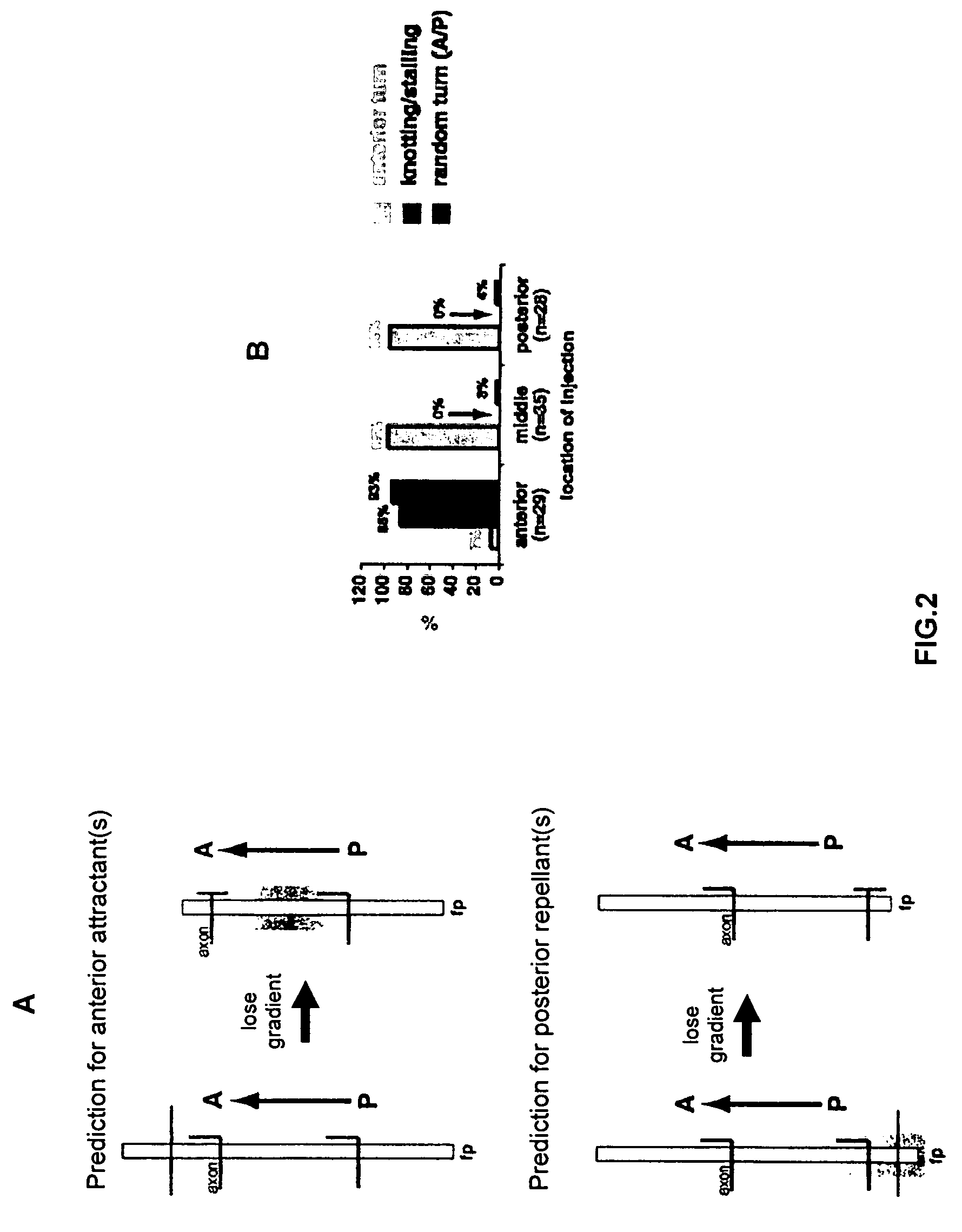
The A-P Guidance Cue(s) is Attractive

[0229]To address whether the A-P guidance cue is attractive or repulsive, DiI was focally injected into the dorsal spinal cord close to the anterior end, in the middle and close to the posterior end of the long explants (3 mm or longer). The axons in the middle and close to the posterior end of the explants were found to always project anteriorly, whereas the axons close to the anterior end almost always make mistakes: they either stall after they cross the midline or they project both anteriorly and posteriorly after midline crossing, or sometimes only posteriorly. The results were quantified using the same criteria as shown in FIG. 1E. The quantification is shown in FIG. 2B. The axons close to the anterior end of the explants behave similarly to those in the short explants (0.5 mm), whereas the axons in the middle and posterior part of the explants behave normally. These results are consistent with the possibility that a gradient of an attracti...


PUM

PropertyMeasurementUnit
concentrationaaaaaaaaaa

Abstract

Methods and compositions for modulating growth of a neuron with a Wnt, a Wnt-like substance, and / or a chemical compound affecting a Wnt signaling pathway are disclosed. Also disclosed are methods for identifying a substance that modulates growth of a neuron by obtaining a candidate substance and contacting the candidate substance with the neuron are disclosed and methods for modulating growth of a neuron in a subject using a Wnt, a Wnt-like substance, and / or a chemical compound affecting a Wnt signaling pathway. The Wnt, Wnt-like substance, and / or chemical compounds affecting a Wnt signaling pathway can be delivered to the subject using gene therapy techniques. Also disclosed are pharmaceutical compositions for modulating growth of a neuron in a mammal that include a Wnt or a Wnt-like substance. Methods and compositions for inhibiting growth of a neuron are also disclosed.

Description

CROSS-REFERENCE TO RELATED APPLICATIONS[0001]This application claims the benefit of U.S. Provisional Application No. 60 / 470,913 filed May 15, 2003.BACKGROUND OF THE INVENTION[0002]1. Field of the Invention[0003]The present invention relates generally to the fields of molecular biology, cell biology, pharmacology, developmental neuroscience, neurology, neurosurgery and regenerative biology. More particularly, it concerns methods and compositions for modulating regeneration of a nerve cell using a Wnt, a Wnt-like substance, and / or a chemical compound affecting a Wnt signaling pathway. It also concerns methods and compositions for inhibiting growth of a neuron using inhibitors of neuronal growth that act via the Wnt signaling pathways, such as a Secreted Frizzled-Related Protein (sFRP), sFRP-like substance, Ryk, or Ryk-like substance.[0004]2. Description of Related Art[0005]The central nervous system (CNS) is connected by ascending sensory pathways and descending motor or regulatory pa...

Claims


Application Information

Patent Timeline
Patent Type & Authority Patents(United States)
IPC IPC(8): A61K39/00A61K39/395C12N5/00C12N5/06C12N5/08A61K31/00A61K38/17A61K38/18A61P25/00A61P25/28G01N33/68
CPCA61K31/00A61K38/1709A61K38/177A61K38/18A61K38/45G01N33/5058A61K2300/00A61K48/00A61P25/00A61P25/28
Inventor ZOU, YIMIN
Owner UNIVERSITY OF CHICAGO
Why Eureka
Try Eureka