Non-invasive method and apparatus to detect and monitor early medical shock, and related conditions

a non-invasive, medical shock technology, applied in the direction of optical radiation measurement, instruments, spectrometry/spectrophotometry/monochromators, etc., can solve the problems of reducing the availability of blood for other vital organs, affecting the metabolism of organs, and affecting the perfusion of other vital organs

Inactive Publication Date: 2009-01-27
CARDIOSENSE LTD
View PDF10 Cites 8 Cited by
  • Summary
  • Abstract
  • Description
  • Claims
  • Application Information

AI Technical Summary

Benefits of technology

[0045]Optionally, potentially false color readings originating from capillary damage due to repeated testing of a skin area may be avoided by ...

Problems solved by technology

Unless shock is promptly treated, this deprivation of blood may give rise to a disturbance in the metabolism of the organ with a resultant damage thereto.
But it is otherwise irreversible and may lead to the death of the patient.
Such losses may be due to dehydration, vomiting, diarrhea, burns, or because of the abusive use of diuretics.
Many cases of bleeding are occult (e.g. slow internal bleeding), and therefore can not be diagnosed early.
The sequestration and pooling of blood in various vascular compartments reduces the availability of blood for the perfusion of other vital organs.
Obstruction to cardiac filling shock takes place when this filling activity is lessened or arrested by a massive pulmonary embolism, or by space-occupying lesions.
Neurogenic shock results from a severe spinal cord injury, or from a massive intake of a depressant drug, causing a loss of vasometric tone.
This reduction in skin perfusion is the result of a profound vasoconstriction of the skin tissue arterioles, which leads to decreased capillary flow, and a resultant poor perfusion to the skin.
When applying pressure onto a specific skin area, the capillaries below the depressed area collapse and blood is blanched therefrom, thereby causing the skin color in the depressed skin area to ...

Method used


Image

  • Non-invasive method and apparatus to detect and monitor early medical shock, and related conditions
  • Non-invasive method and apparatus to detect and monitor early medical shock, and related conditions
  • Non-invasive method and apparatus to detect and monitor early medical shock, and related conditions

Examples


first embodiment

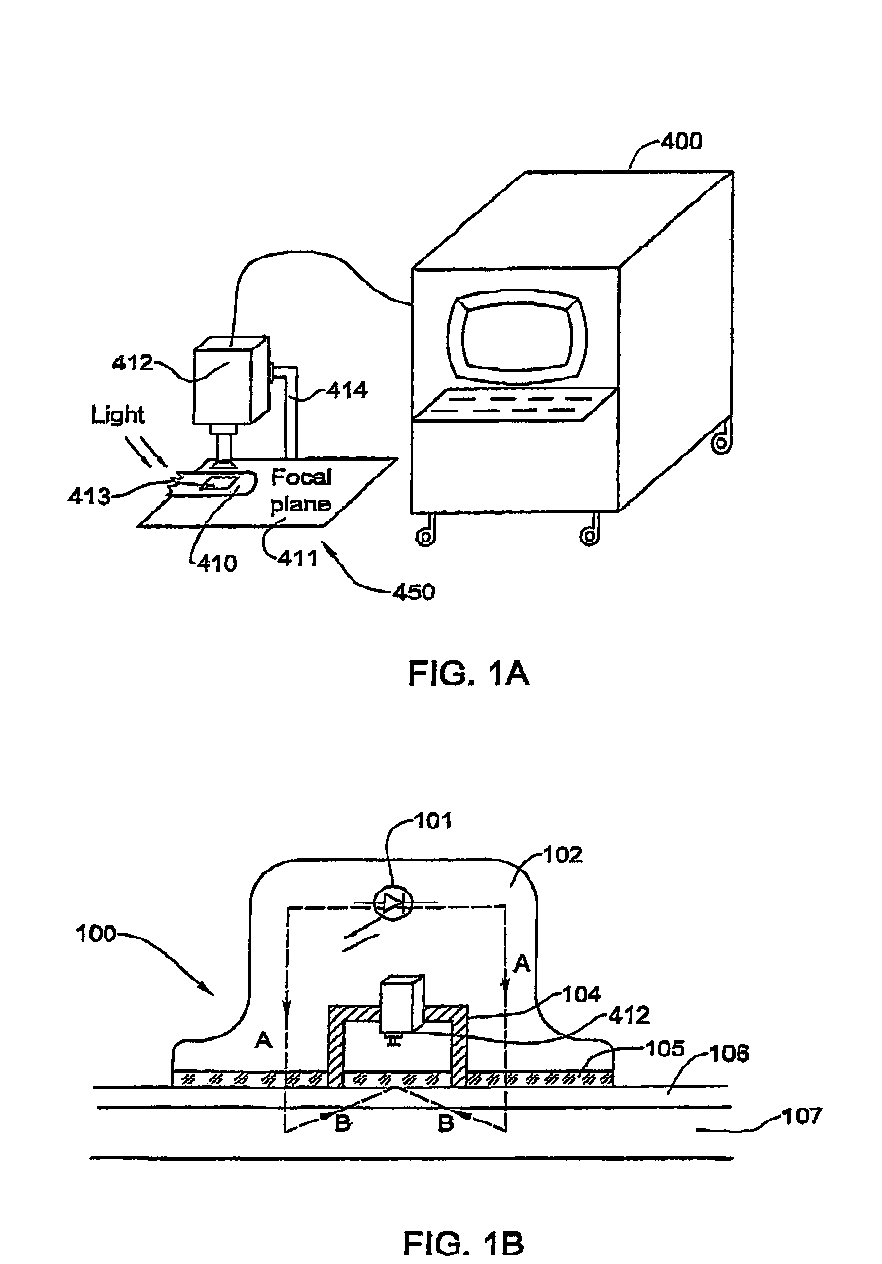
[0059]Schematically illustrated in FIG. 1A is a CFT instrument 450 adapted to diagnose a shock-related condition in a patient by measuring capillary filling time and rate.

[0060]Instrument 450 includes a camera 412, such as a color video camera, fixed in place by a holder 414 above a rigid table surface 411 on which an appendage 410 of a patient rests. This appendage may for example be the patient's finger. The position of camera 412 is adjusted so that the skin area 413 viewed by the camera for purposes of CFT measurement, is in or is close to the focal plane of the lens. Pressure may be applied to and released from skin area 413 manually or by using mechanical apparatus which may be automatically controlled.

[0061]Skin area 413 is illuminated by background light, and light reflected from the surface of this area is received in the lens of camera 412. A minimal illumination level of 0.2 lux is sufficient for most currently-available modern cameras for color discrimination. Camera 412...

second embodiment

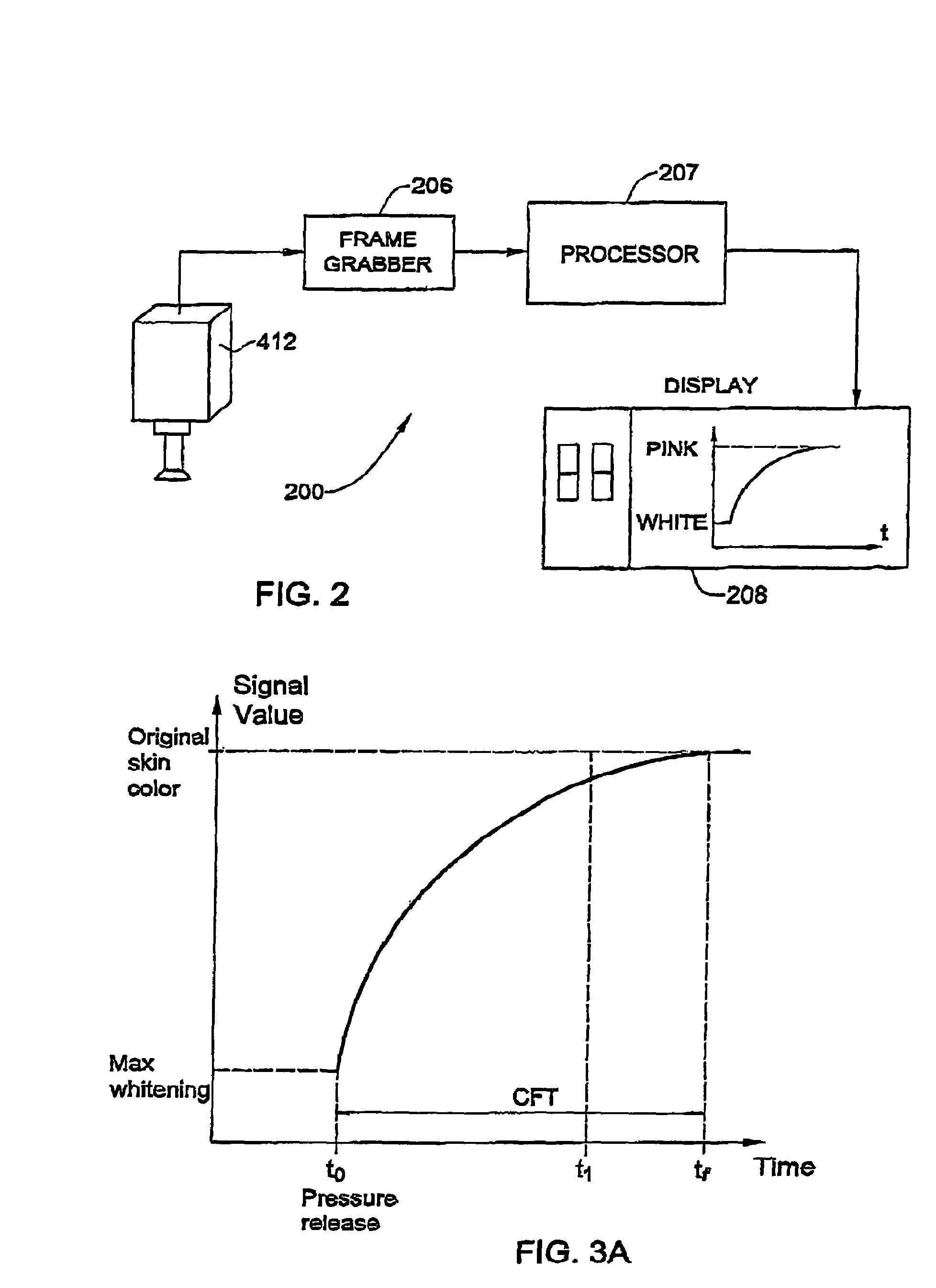
[0086]This embodiment of a CFT diagnostic instrument differs from the instrument shown in FIG. 1 mainly in the nature of its skin color sensor. However, in all other respects it operates in the same manner as does the first embodiment.

[0087]FIG. 5. Schematically illustrates the structure of a skin color sensing device 500 for the diagnosis of a shock-related state in a patient by measure the capillary filling time and rate according to the second embodiment of the invention. Device 500 includes a continuous (non-modulated) or a pulsating (modulated) light source 501, such as a Light Emitting Diode (LED) driven by a rectangular voltage pulse generator at a predetermined frequency fo. Light source 501 is enclosed in a light-reflecting external housing 502 having an opening in its bottom side so that most of the light radiation emitted from light source 501 is directed toward the bottom side in one direction “A”. External housing 502 has within it an opaque internal housing 504 contain...

third embodiment

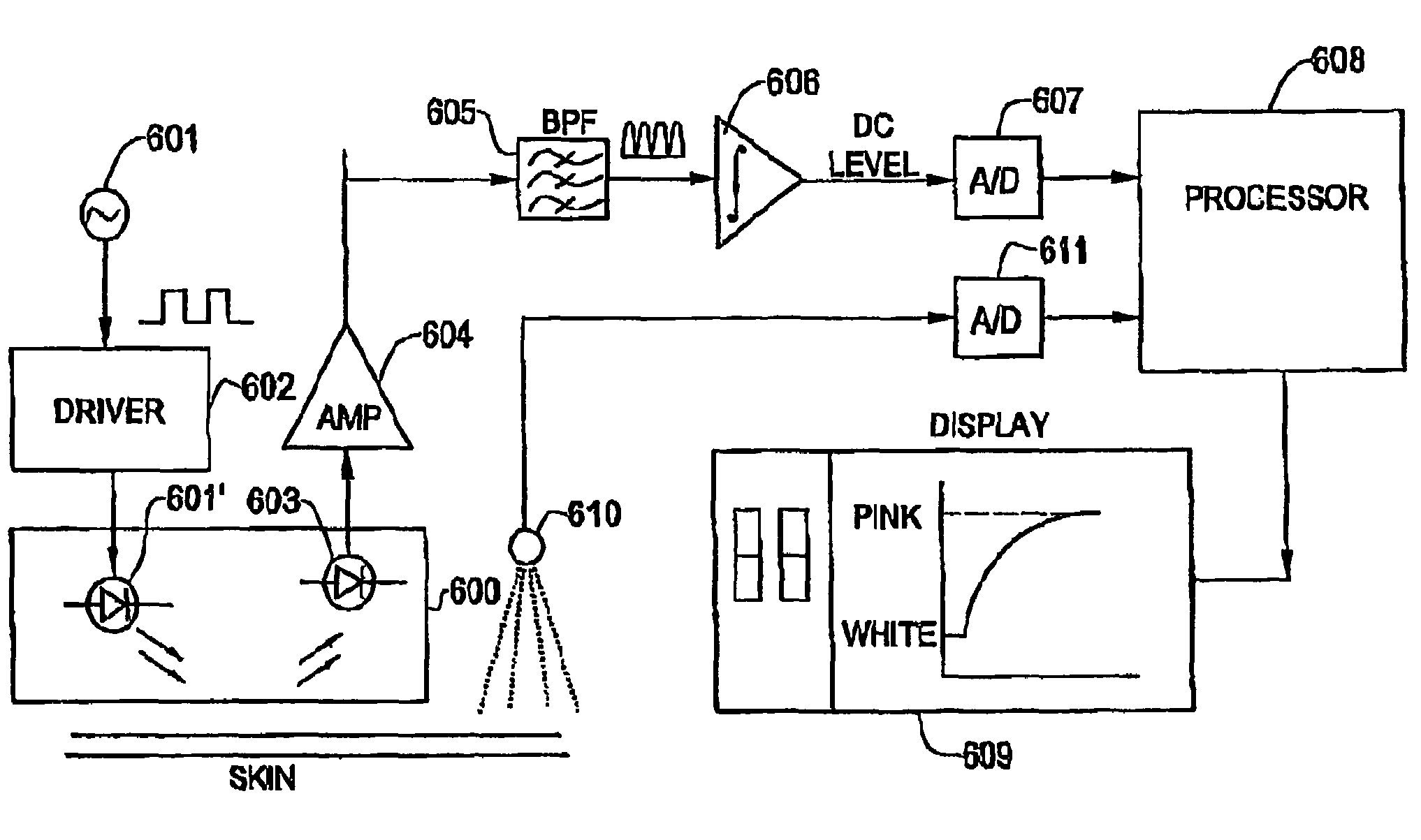
[0095]The third embodiment is substantially similar to the second embodiment, as described herein, with the following differences, mutatis mutandis. FIG. 9 is a block diagram of an apparatus 700 in the third embodiment for diagnosing a shock-related state in a patient by measuring capillary filling time and rate. Apparatus 700 comprises a constant source 712 operated at a DC voltage. The output of source 712 is fed into a driver 702 which provides energy to power light source 701 to emit non-modulated, continuous light. Light reflected from the skin is converted by light sensor 703 to a corresponding electrical signal. This signal is fed into an amplifier 704 operating at near-DC frequency band to increase the amplitude of the electrical signal.

[0096]Light sensor 703 is may be sensitive to the full color spectrum, or alternatively most sensitive to light radiation to a particular range of wavelengths, for example between red and infra-red in the color spectrum The sensor 703 may als...


PUM


Abstract

A diagnostic medical instrument adapted to determine whether a patient is suffering from a pre-shock, shock, or shock-related condition. The instrument is used in a capillary filling time CFT test procedure in which a skin area in the patient which overlies blood-filled capillaries which normally display a pink color is depressed to expel blood from the capillaries and impart white color to the skin at which point the pressure is released to permit blood to flow back into the capillaries and cause the skin to regain its pink color. The instrument includes a color sensor trained on the skin area and responsive to light reflected therefrom to produce a first signal at the point in time the skin color tuns from pink to white and to later produce a second signal at the point in time at which the skin color has turned from white to pink. The time elapsing between the first and second signals is measured to provide a CFT index indicative of the patient's condition.

Description

FIELD OF THE INVENTION[0001]This invention relates generally to the diagnosis of medical shock-related conditions and to instruments for this purpose. More particularly, the invention relates to methods and apparatus for the non-invasive detection of pre-shock, shock and shock-related conditions (other related causes of cardio-pulmonary distress), and for assisting in a patient's recovery from these conditions by monitoring changes in capillary flow in skin areas of peripheral body organs.BACKGROUND OF THE INVENTION[0002]The normal skin color at most sites on the human body is generally pink. Skin color depends on the amount of blood flowing in the capillaries through which blood flows from the arterioles to the venules. The present invention resides in non-invasive invasive detection of hemodynamic changes in the skin arteriolar-capillary flow during states of pre-shock, shock and cardiopulmonary distress. These changes are indicative of a reduction in blood delivery to an organ of...

Claims


Application Information

Patent Timeline
IPC IPC(8): A61B5/145A61B5/103
CPCA61B5/0059A61B5/1032A61B5/441
Inventor SHANI, HAIMSHAVIT, ITAI
Owner CARDIOSENSE LTD
Why Eureka
Try Eureka