
Diagnostic methods

a technology of helicobacter pylori and diagnostic methods, which is applied in the field of new diagnostic methods for helicobacter pylori infection, can solve the problems of less suitable use for evaluation and follow-up of treatment and cure, relying on the detection of specific antibodies against i>h. pylori, and the relative long time needed for reliable test, etc., to achieve reliable detection of h and increase the sensitivity of the assay

Inactive Publication Date: 2009-06-09
BIONAVIS
View PDF76 Cites 4 Cited by
  • Summary
  • Abstract
  • Description
  • Claims
  • Application Information

AI Technical Summary

Benefits of technology

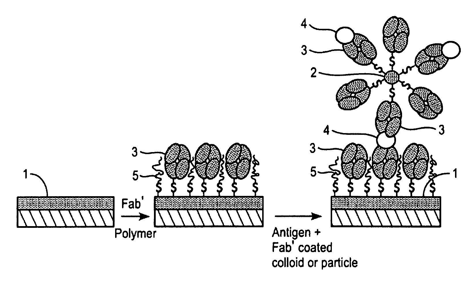
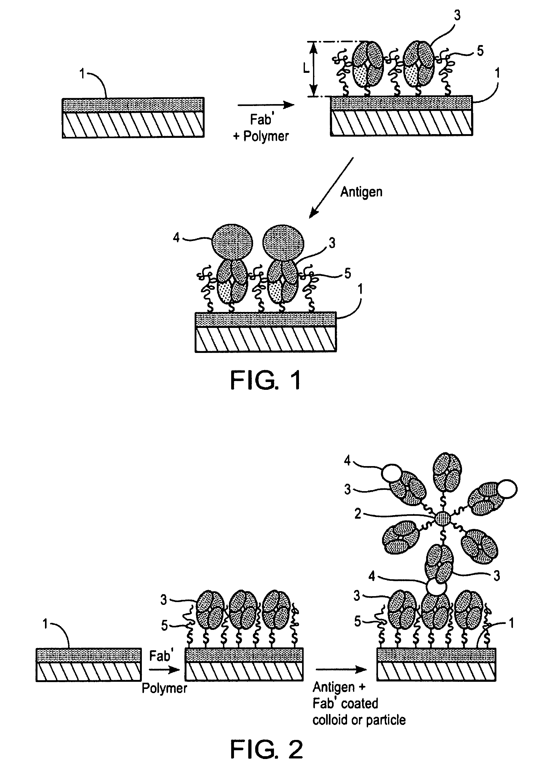
[0013]It was unexpectedly found that it is possible to non-invasively detect the presence or the absence of Helicobacter pylori in a biological sample employing methods based on the use of a highly specific and sensitive biosensor and thereby meet the objects of the present invention. The biosensor useful in the present invention comprises a carrier substrate, onto which specific antibodies against H. pylori or antigen-binding fragments thereof have been directly attached together with biomolecule-repellent molecules, which cover the surface between the immobilized antibodies or the antibody fragments. These biomolecule-repellent molecules efficiently prevent the undesired non-specific binding of the analyte and the contaminant (bio)molecules present in the biological sample and highly increase the sensitivity of the assay. The use of such a biosensor affords reliable detection of H. pylori antigens and / or metabolites produced by the bacterium in any biological specimen.

Problems solved by technology

The determination of antibodies against H. pylori suffer from several drawbacks, such as the strong dependence of the antigen preparation which is used to capture the antibodies, cross reactions of antibodies from related bacterial species, and the relatively long time needed for reliable test results.
The accuracy of the so-called “office-based” or “near-patient” tests offered for use in doctor's offices is poorer than that of conventional laboratory assays.
Importantly, these assays relying on the detection of specific antibodies against H. pylori are less suitable for use in the evaluation and follow-up of the treatment and cure, since the elevated antibody levels maintain for a long period of time after the treatment and cure of the infection.
However, the conventional immunoassays rely on a marker molecule such as a radioactive label, an enzyme label, a fluorescent label or chemiluminescent label, and are laborious and time-consuming, since several incubation, washing and separation steps are needed before the actual detection.
Additionally, for a reliable performance, they require a skilled personnel and rather expensive apparatuses.
The sample, especially a faecal sample, may also represent a problem.
Many patients find the collection of one faecal sample, let alone the collection of several faecal samples necessary for the follow-up, unpleasant and not hygienic, and their compliance to the treatment may suffer.
Similarly, the personnel may dislike the handling of the faecal specimen and the preparation of samples for such assays due to the inherent infection risk.

Method used


Image

  • Diagnostic methods
  • Diagnostic methods
  • Diagnostic methods

Examples


example 1

A General Procedure for the Preparation of the Carrier Substrate of the Invention

[0042]For the preparation of the carrier substrate, Fab′-fragments were first prepared from a specific monoclonal anti-H. pylori antibody as follows. First, F(ab′)2 fragments were prepared with ImmunoPure F(ab′)2 Preparation Kit (PIERCE, USA) from monoclonal anti-H. pylori antibodies, such as mono-clonal anti-H. pylori antibodies clones 7101 and 7102 (Medix Biotechnica, Kauniainen, Finland). Other known commercial kits and methods can equally be used. Then the F(ab′)2 fragments were split into Fab′ fragments with dithiothreitol (DTT, Merck) in a HEPES / EDTA buffer containing 150 mM NaCl, 10 mM HEPES, 5 mM EDTA, pH 6.0, typically over night in a microdialysis tube as described by Ishikawa [Ishikawa, E., J. Immunoassay 4 (1983) 209-320]. Briefly, F(ab′) 2 fragments at a concentration of 0.2-0.5 mg / ml were mixed with HEPES / EDTA buffer and 6.25 mM DTT solution in a microdialysis tube. The dialysis tube was i...

example 2

A General Procedure for the Measurement of H. pylori Antigens with SPR

[0045]H. pylori antigens can be measured in a biological sample with the following general SPR procedure. The surface of carrier substrate prepared as described in Example 1 is rinsed with PBS, pH 7.2. A negative sample (blank) is run at first. The negative sample used depends on the biological sample to be measured. Thus when, for example, a stool sample is to measured, the negative sample is a stool sample negative for H. pylori, when a urine sample is to be measured, the negative sample is a urine sample obtained from a subject without H. pylori infection, and when a serum sample is to be measured, the negative sample is a serum sample obtained from a subject without H. pylori infection. Then surface of the carrier substrate is sequentially brought into contact with the standard solutions, positive and negative controls and the samples by filling the flow cell of the measuring device for 10 minutes each with th...

example 3

Preparation of a Standard Curve and the Reproducibility of the Measurement

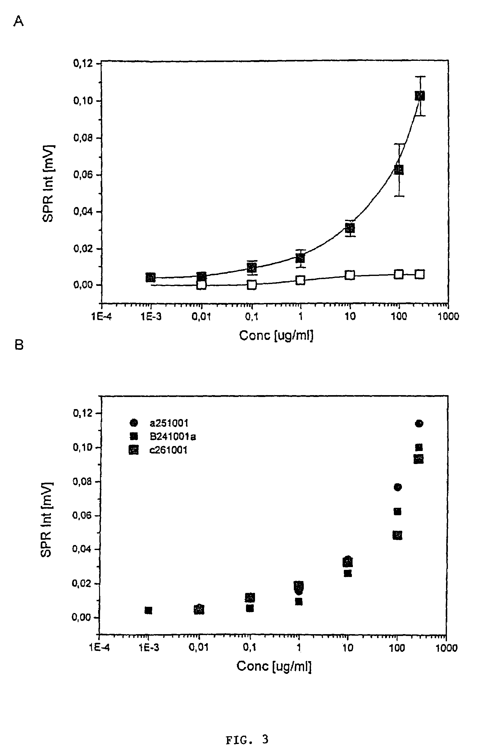
[0046]The H. pylori antigen, which was used as a standard, was extracted from the bacterial mass of H. pylori strain ATCC 49503 using the glycine-acid extraction procedure described by Rautelin and Kosunen [J. Clin. Microbiol. 25 (10) (1987) 1944-1951]. The protein concentration was determined with Bradford assay [Bradford, Anal. Biochem. 72 (1976) 248]. The standards were diluted in 0.1 M PBS, pH 7.2, to concentrations of 0.001, 0.01, 0.1, 1, 10, 100 and 270 μg / ml and run following the general procedure described in Example 2. Three separate measurements were made on consequent days to analyse the reproducibility.

[0047]The results are shown in FIG. 3A. The standard curve shown in FIG. 3A shows that the response is directly comparable with the antigen concentration on a semilogarithmic scale. Table 1 shows the standard deviations from three separate measurements of the standard curve on consequent days (FIG. 3...


PUM

PropertyMeasurementUnit
pHaaaaaaaaaa
pHaaaaaaaaaa
concentrationaaaaaaaaaa

Abstract

The present invention relates to novel methods for the diagnosis of Helicobacter pylori infection. Specifically, the present invention relates to novel non-invasive methods for the detection of the presence or absence of a Helicobacter pylori antigen or a metabolite produced by the bacterium in a biological sample with a biosensor-based measurement. The present invention also related to the use of a biosensor containing specific antibodies against H. pylori or antigen-binding fragments thereof immobilized thereto together with biomolecule-repellent polymers preventing the non-specific binding. The invention also relates to test kits useful in the methods.

Description

FIELD OF THE INVENTION[0001]The present invention relates to novel methods for the diagnosis of Helicobacter pylori infection. Specifically, the present invention relates to novel non-invasive methods for the detection of the presence or absence of a Helicobacter pylori antigen or a metabolite produced by the bacterium in a biological sample with a biosensor-based measurement. The present invention also related to the use of a biosensor containing specific antibodies against H. pylori or antigen-binding fragments thereof immobilized thereto together with biomolecule-repellent polymers preventing the non-specific binding. The invention also relates to test kits useful in the methods.BACKGROUND OF THE INVENTION[0002]Helicobacter pylori is a curved gram-negative bacterium found in the upper gastrointestinal tract of humans. Since the first isolation of the bacterium in 1982, a huge amount of evidence has accumulated on the association of H. pylori with various gastric disorders, includ...

Claims


Application Information

Patent Timeline
Patent Type & Authority Patents(United States)
IPC IPC(8): C12M1/34C07K14/205G01N21/27G01N33/543G01N33/553G01N33/569
CPCG01N33/54393G01N33/56922Y10S435/808Y10S436/805Y10S436/806
Inventor KLEIMOLA, VESAEEROLA, ERKKIVIANDER, MARKKU
Owner BIONAVIS
Learn More