Protein subcellular localization assays using split fluorescent proteins

a technology of fluorescent proteins and subcellular localization, which is applied in the direction of instruments, peptides, organic chemistry, etc., can solve the problems of misfolding of the fused protein, interference with protein processing and/or intracellular transport, and limited information provided by methods

a technology of fluorescent proteins and subcellular localization, which is applied in the direction of instruments, peptides, organic chemistry, etc., can solve the problems of misfolding of the fused protein, interference with protein processing and/or intracellular transport, and limited information provided by methods

US7585636B2Active Publication Date: 2009-09-08TRIAD NAT SECURITY LLC

Image

  • Protein subcellular localization assays using split fluorescent proteins
  • Protein subcellular localization assays using split fluorescent proteins
  • Protein subcellular localization assays using split fluorescent proteins

Examples


example 1

Complementation of Split GFP in the Cytoplasm—Detection of Cytoplasmic Proteins

Expression of Fluorescent Tagged Proteins:

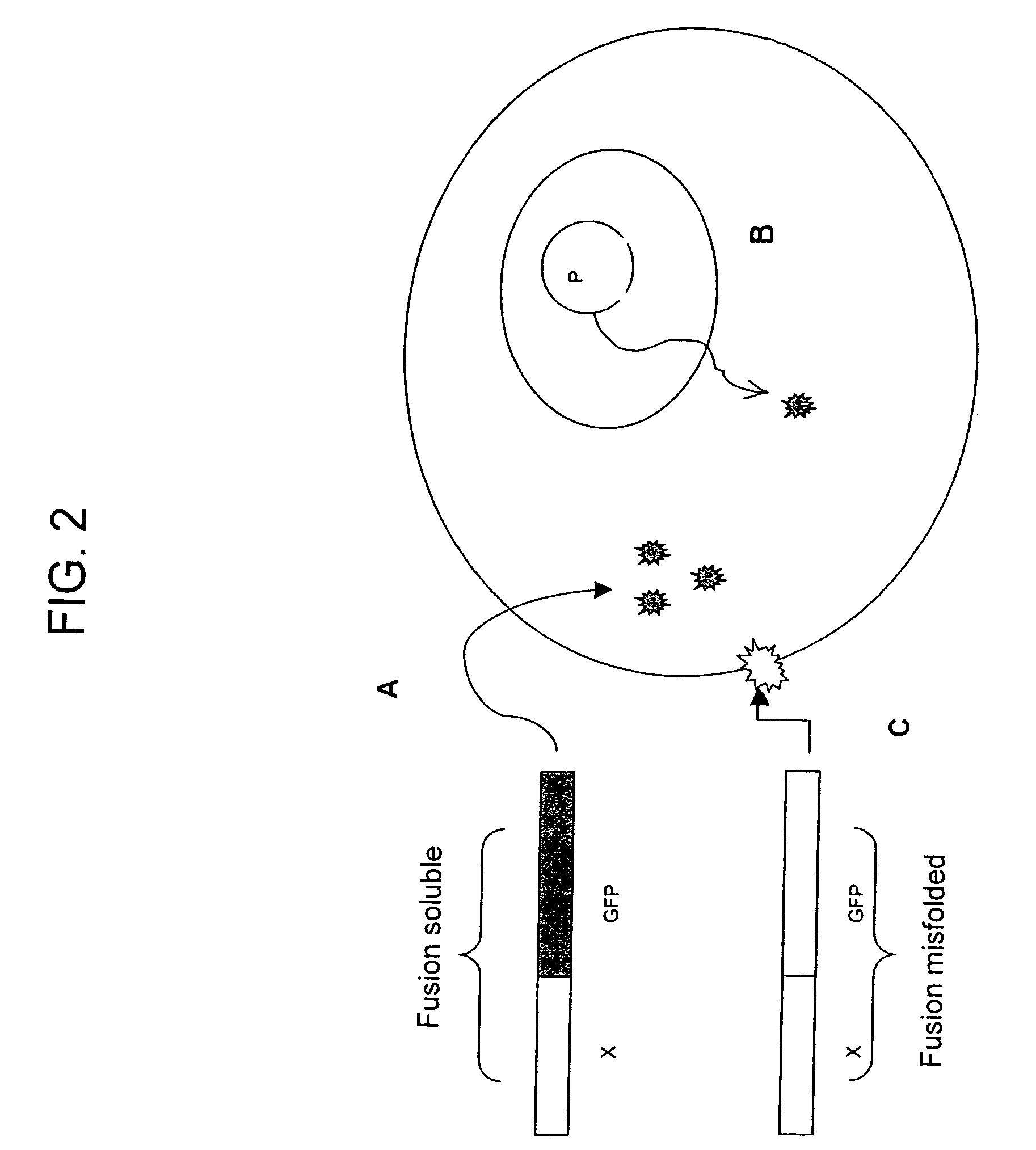
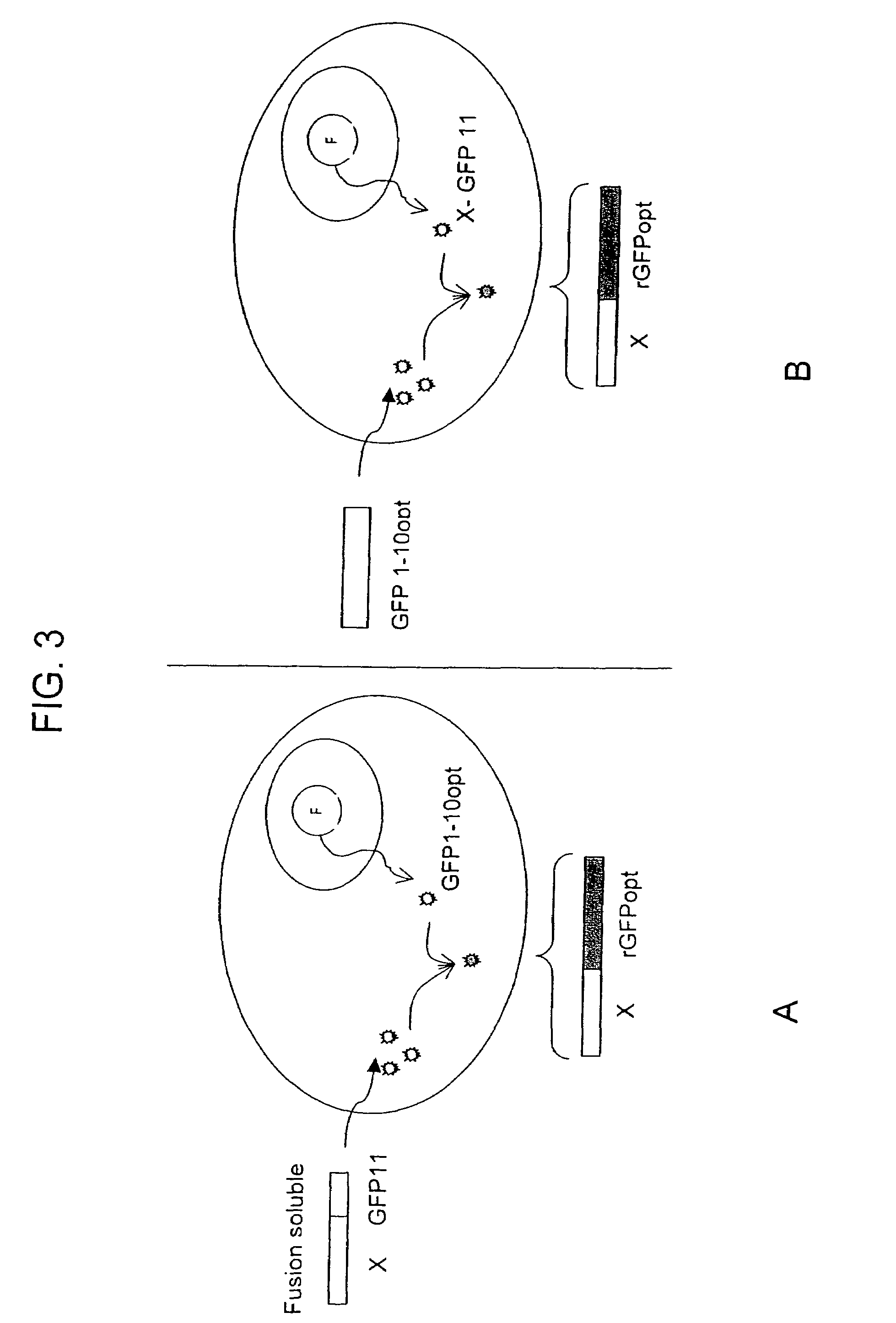
[0107]A test protein, in this example, the soluble p. aerophilum sulfite reductase gamma subunit (SR), is tagged with a single beta-strand microdomain of a fluorescence protein, i.e. GFP s11 opt, and a complementary “assay” fragment of this fluorescent protein (i.e. GFP s1-10) is expressed from N-6HIS pET 28 vectors (Novagen, Madison, Wis.). 500 ml cultures of BL21 (DE3) expressing each construct are grown to OD600 ˜0.5, and induced with 1 mM IPTG for 4 h at 37° C. The culture pellets are resuspended in 10 ml TNG and sonicated. The soluble fractions are loaded onto Talon resin purification beads (TALON, Clontech, Palo Alto, Calif.). After two washes with excess TNG buffer and one wash in TNG supplemented with 5 mM imidazole, the protein is eluted with 150 mM imidazole in TNG buffer. Proteins are dialyzed in PBS buffer (mMNaCl, mMKCl, mMNa2HPO4, mMKH2PO4), quantifi...

example 2

Complementation of Split GFP in Specialized Compartments

Cloning of Localization Sequences in Fusion with SR-s11 and GFP1-10

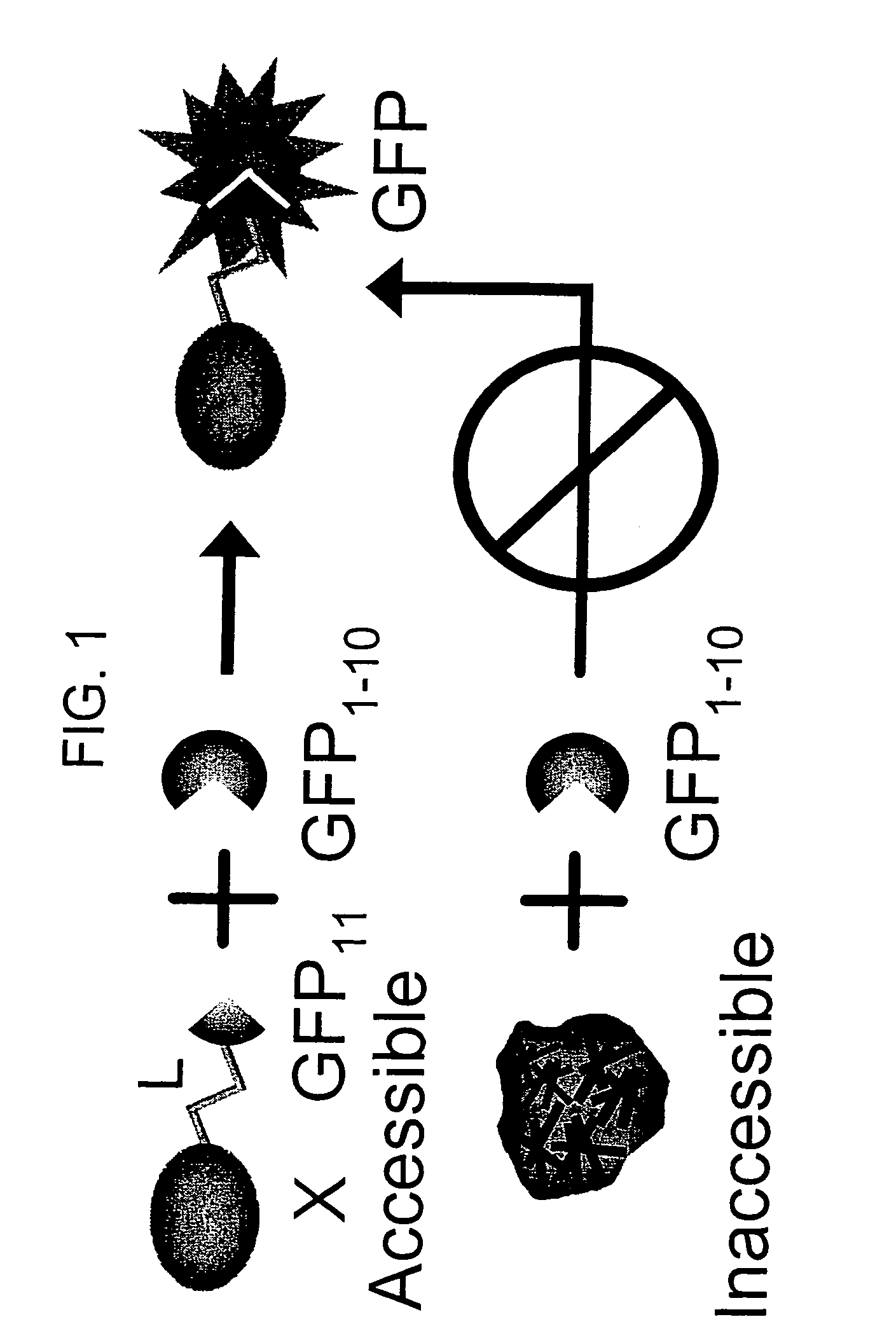
[0114]If both fragments contains a sequence that targets the gene of interest in a specialized compartment, then the two tagged split-GFP fragments will be targeted to this particular location and will be able to complement in the compartment.

[0115]Various subcellular localization signal sequences or tags are known and / or commercially available. These tags are used to direct split-fluorescent protein fragments to particular cellular components or outside of the cell. Mammalian localization sequences capable of targeting proteins to the nucleus, cytoplasm, plasma membrane, endoplasmic reticulum, golgi apparatus, actin and tubulin filaments, endosomes, peroxisomes and mitochondria are known.

[0116]Detailed protocols for localization in the nucleus, mitochondria and endoplasmic reticulum follow.

[0117]Localization in mitochondria: The mitochondrial targeting sequence...


PUM

PropertyMeasurementUnit
emission wavelengthsaaaaaaaaaa
pHaaaaaaaaaa
concentrationaaaaaaaaaa

Abstract

The invention provides protein subcellular localization assays using split fluorescent protein systems. The assays are conducted in living cells, do not require fixation and washing steps inherent in existing immunostaining and related techniques, and permit rapid, non-invasive, direct visualization of protein localization in living cells. The split fluorescent protein systems used in the practice of the invention generally comprise two or more self-complementing fragments of a fluorescent protein, such as GFP, wherein one or more of the fragments correspond to one or more beta-strand microdomains and are used to “tag” proteins of interest, and a complementary “assay” fragment of the fluorescent protein. Either or both of the fragments may be functionalized with a subcellular targeting sequence enabling it to be expressed in or directed to a particular subcellular compartment (i.e., the nucleus).

Description

RELATED APPLICATION[0001]This patent application claims the benefit of the filing date of U.S. Provisional patent application No. 60 / 633,488, filed Dec. 4, 2004. The subcellular localization assays of the invention utilize split-fluorescent and split-chromophoric protein systems, which are described in co-owned, co-pending U.S. patent application Ser. No. 10 / 973,693, entitled “SELF-ASSEMBLING SPLIT-FLUORESCENT PROTEIN SYSTEMS”, filed Oct. 25, 2004 and hereby incorporated by reference in its entirety.STATEMENT AS TO RIGHTS TO INVENTIONS MADE UNDER FEDERALLY SPONSORED RESEARCH OR DEVELOPMENT[0002]This invention was made with government support under Contract No. W-7405-ENG-36 awarded by the United States Department of Energy to The Regents of The University of California. The government has certain rights in this invention.BACKGROUND OF THE INVENTION[0003]Understanding the intracellular transport and localization of proteins is important because many processes in biology, including tr...

Claims


Application Information

Patent Timeline
08 Sep 2009
Publication
US7585636B2
IPC
G01N33/567; C12Q1/68; G01N33/53
CPC
G01N33/5005; G01N33/5041; G01N33/6842; G01N33/582; G01N33/6803; G01N33/542
Inventors
WALDO, GEOFFREY S.; CABANTOUS, STEPHANIE