Biglycan and related therapeutics and methods of use

a biglycan and related technology, applied in the field of biglycans, can solve the problems of affecting the survival rate of patients, and unclear how agrin stimulates musk, and achieves the effect of less “leaky” and longer cell life span

Inactive Publication Date: 2009-11-03
BROWN UNIV RES FOUND INC
View PDF5 Cites 12 Cited by
  • Summary
  • Abstract
  • Description
  • Claims
  • Application Information

AI Technical Summary

Benefits of technology

[0013]The invention provides methods and compositions for stabilizing dystrophin-associated protein complexes (DAPCs) on the surface of a cell. Stabilizing DAPC complexes on cell membranes allows membranes to be less “leaky” and thus, provides a longer life span to cells. The invention also provides methods for activating a postynaptic membrane, such as to render the membrane more sensitive to an incoming signal from a neural cell (e.g., at a neuromuscular junction). Activating a postsynaptic membrane may comprise stimulating aggregation of AChR on the cell membrane and / or activating MuSK, such as by phosphorylation.
[0018]Moreover, as is apparent from the present disclosure, mimetopes of the subject biglycan peptides can be provided. Such peptidomimetics can have such attributes as being non-hydrolyzable (e.g., increased stability against proteases or other physiological conditions which degrade the corresponding peptide), increased specificity and / or potency, and increased cell permeability for intracellular localization of the peptidomimetic. For illustrative purposes, peptide analogs of the present invention can be generated using, for example, benzodiazepines (e.g., see Freidinger et al. in Peptides: Chemistry and Biology, G. R. Marshall ed., ESCOM Publisher: Leiden, Netherlands, 1988), substituted gama lactam rings (Garvey et al. in Peptides: Chemistry and Biology, G. R. Marshall ed., ESCOM Publisher: Leiden, Netherlands, 1988, p123), C-7 mimics (Huffman et al. in Peptides: Chemistry and Biologyy, G. R. Marshall ed., ESCOM Publisher: Leiden, Netherlands, 1988, p. 105), keto-methylene pseudopeptides (Ewenson et al. (1986) J Med Chem 29:295; and Ewenson et al. in Peptides: Structure and Function (Proceedings of the 9th American Peptide Symposium) Pierce Chemical Co. Rockland, Ill., 1985), β-turn dipeptide cores (Nagai et al. (1985) Tetrahedron Lett 26:647; and Sato et al. (1986) J Chem Soc Perkin Trans 1:1231), β-aminoalcohols (Gordon et al. (1985) Biochem Biophys Res Commun126:419; and Dann et al. (1986) Biochem Biophys Res Commun 134:71), diaminoketones (Natarajan et al. (1984) Biochem Biophys Res Commun 124:141), and methyleneamino-modifed (Roark et al. in Peptides: Chemistry and Biology, G. R. Marshall ed., ESCOM Publisher: Leiden, Netherlands, 1988, p134). Also, see generally, Session III: Analytic and synthetic methods, in Peptides: Chemistry and Biology, G. R. Marshall ed., ESCOM Publisher: Leiden, Netherlands, 1988)

Problems solved by technology

Perturbations of these specializations, such as the decrease in the number of functional AChRs seen in myasthenia gravis, can lead to debilitating and often fatal clinical outcomes (Oosterhuis, et al (1992) Neurology &Neurosurgery 5: 638).
However, agrin does not bind to MuSK and it is unclear how agrin stimulates MuSK.
However, MuSK phosphorylation is necessary but not sufficient for agrin-induced AChR clustering.

Method used


Image

  • Biglycan and related therapeutics and methods of use
  • Biglycan and related therapeutics and methods of use
  • Biglycan and related therapeutics and methods of use

Examples


example 1

Characterization of a Dystroglycan-Binding Protein, DAG-125

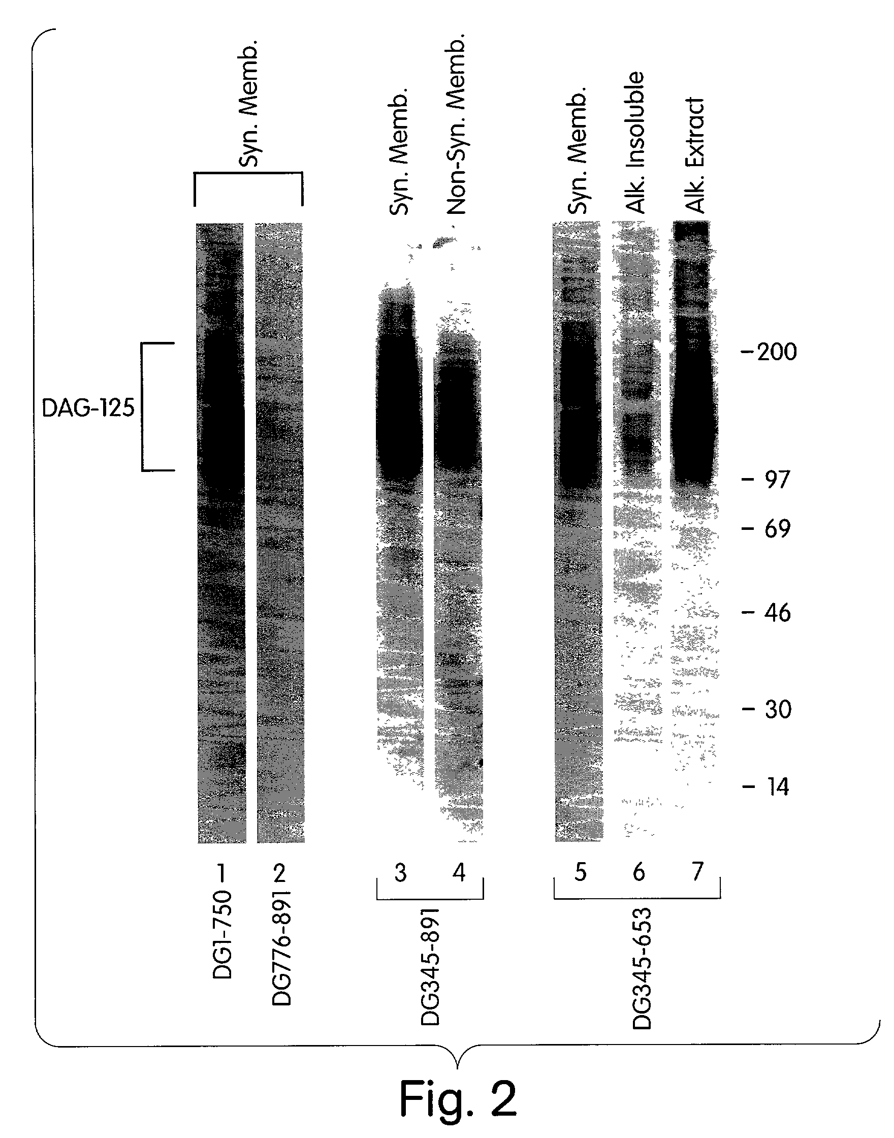
[0304]This Example describes the identification of a dystroglycan-binding protein, termed DAG-125.

[0305]In order to identify novel dystroglycan binding partners, a ligand blot overlay assay, was developed as follows. Postsynaptic and non-synaptic membrane fractions from Torpedo electric organ were prepared as previously described (Bowe, et al. (1994) Neuron. 12: 1173). All handling of membranes and protein was performed at 4° C.

[0306]Membrane proteins were separated by SDS-PAGE (5-15% gradient gel), and transferred to nitrocellulose. To detect dystroglycan binding proteins, the nitrocellulose was rinsed and blocked for 3 hr in Hank's Balanced Salt Solution containing 1 mM CaCl2, 1 mM MgCl2, 1% bovine serum albumin, 1% Nonfat Dry Milk, 1 mM DTT, 10 mM HEPES, pH 7.4, and was then incubated overnight in the same buffer containing 35S-methionine-labelled dystroglycan fragments produced by in vitro transcription / translation as fo...

example 2

Association Between α-dystroglycan and DAG-125

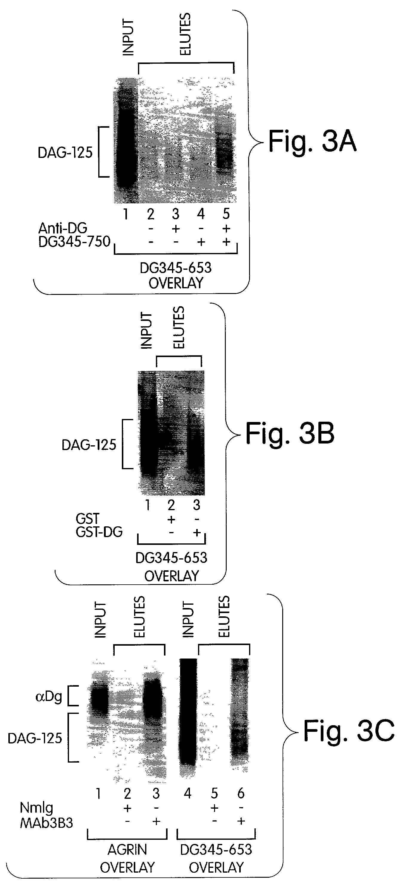
[0312]This Example demonstrates that DAG-125 associates with in vitro-translated α-dystroglycan, bacterially produced GST-α-dystroglycan fusion protein and native α-dystroglycan in solution.

[0313]DAG-125 was solubilized by alkaline-treatment, and neutralized, as described above, and incubated with column matrices and recombinant or native dystroglycan as indicated in FIG. 3. The input material and eluates from the beads were analyzed by ligand blot overlay assay for the presence of DAG-125 (35S-DG345-653 as probe) or native α-dystroglycan (agrin overlay, see Example 1).

[0314]FIG. 3A shows DAG-125 incubated with goat anti-mouse Ig-conjugated agarose beads in the presence or absence of in vitro translated dystroglycan polypeptide (DG345-750) and / or anti-dystroglycan monoclonal antibody (NCL-β-DG; Novocastra, Newcastle-on-Tyne, UK). The results indicate that DAG-125 co-precipitated with dystroglycan plus anti-dystroglycan antibody (lane 5),...

example 3

Localization of the DAG-125 Binding Domain of α-dystroglycan

[0318]This Example describes that the DAG-125 binding domain of α-dystroglycan is contained in an approximately 150 amino acid carboxyl-terminal domain of the protein.

[0319]In order to determine the region of α-dystroglycan that interacts with DAG-125, a panel of dystroglycan fragments were prepared by in vitro translation (FIG. 4) and the ability of each to bind DAG-125 was tested using the ligand blot overlay assay. FIG. 4, which show the results, indicates that DAG-125 binds to the carboxyl-terminal one-third of α-dystroglycan. A small contribution from the middle third of α-dystroglycan is also possible. The ectodomain of β-dystroglycan does not appear to contribute to binding of DAG-125. Moreover, these fragments were produced under conditions in which the polypeptides are not glycosylated. Therefore, carbohydrate side chains on dystroglycan are not necessary for its binding to DAG-125.

[0320]Thus, the major binding dom...


PUM

PropertyMeasurementUnit
molecular weightaaaaaaaaaa

Abstract

The invention provides compositions and methods for treating, preventing, and diagnosing diseases or conditions associated with an abnormal level or activity of biglycan; disorders associated with an unstable cytoplasmic membrane, due, e.g., to an unstable dystrophin associated protein complex (DAPC); disorders associated with abnormal synapses or neuromuscular junctions, including those resulting from an abnormal MuSK activation or acetylcholine receptor (AChR) aggregation. Example of diseases include muscular dystrophies, such as Duchenne's Muscular Dystrophy, Becker's Muscular Dystrophy, neuromuscular disorders and neurological disorders.

Description

CROSS-REFERENCE TO RELATED APPLICATION[0001]This application is a continuation-in-part of U.S. Ser. No. 09 / 715,836 filed 17 Nov. 2000 now U.S. Pat. No. 6,864,236, which claims the benefit of U.S. Provisional Application No. 60 / 166,253, filed 18 Nov. 1999. This application also claims the benefit of U.S. Provisional Application No. 60 / 270,053, filed 20 Feb. 2001. The specifications of each application are specifically incorporated herein.GOVERNMENT GRANTS[0002]This work was made during with Government support under Grants HD23924 and MH53571. awarded by the National Healths Institute. Therefore, the U.S. Government has certain rights in this invention.BACKGROUND OF THE INVENTION[0003]The dystrophin-associated protein complex (DAPC) links the cytoskeleton to the extracellular matrix and is necessary for maintaining the integrity of the muscle cell\plasma membrane. The core DAPC consists of the cytoskeletal scaffolding molecule dystrophin and the dystroglycan and sarcoglycan transmembr...

Claims


Application Information

Patent Timeline
Patent Type & Authority Patents(United States)
IPC IPC(8): A61K38/00A01N37/18A61K39/00A61K45/00A61K38/17G01N33/68
CPCA61K38/1709G01N33/6887G01N2400/12G01N2800/2885G01N2500/04G01N2800/2878G01N2500/02A61P21/00A61P25/00
Inventor FALLON, JUSTIN R.MCKECHNIE, BETHRAFII, MICHAELCREELY, HILLIARYBOWE, MARK A.AMENTA, ALISONMERCADO, MARY LYNNHAGIWARA, HIROKI
Owner BROWN UNIV RES FOUND INC
Why Eureka
Try Eureka