
Method for detecting pathogenic mycobacteria in clinical specimens

a clinical specimen and mycobacteria technology, applied in the field of detection of pathogenic mycobacteria in clinical specimens, can solve the problems of early tuberculosis that goes unrecognized in an otherwise healthy individual, lack of simple, rapid and reliable, and enormous problems for both individual patient management and implementation of appropriate infection control and public health measures

Inactive Publication Date: 2009-12-29
COUNCIL OF SCI & IND RES
View PDF0 Cites 15 Cited by
  • Summary
  • Abstract
  • Description
  • Claims
  • Application Information

AI Technical Summary

Benefits of technology

This approach provides a rapid, safe, and specific detection of pathogenic mycobacteria, improving sensitivity and specificity, reducing false positives and negatives, and enabling the differentiation of species, thus aiding in the management of tuberculosis and co-infections, particularly in HIV patients.

Problems solved by technology

Early tuberculosis often goes unrecognized in an otherwise healthy individual.
The lack of simple, rapid and reliable tests that can specifically detect M. tuberculosis and other causative agents in a clinical specimen poses enormous problems for both individual patient management and implementation of appropriate infection control and public health measures.
However, in a vast majority of cases the sputum smear examination is negative for mycobacteria in the early stages of the disease, and lung changes may not be obvious on a X-ray until several months following infection.
Staining of smear for acid-fast bacilli (AFB) takes less than two hours but lacks sensitivity and may be non-specific in some case.
Culturing of M. tuberculosis and other related organisms is sensitive and specific but is cumbersome and may take 6-12 weeks while culturing on solid media and three to six weeks on liquid media, during which time a patient may become seriously ill and infect other individuals.
Main drawback of these methods is their lack of sensitivity and or specificity (Kadival, et al., 1986; Yenez, et al., 1986).
Serological techniques may be useful in some clinical settings but this approach is limited in general due to poor sensitivity and or specificity.
Major drawback of this method is that this is cumbersome, and takes over 24 hours to complete.
Species specific probes that are used to detect different species sequence in the amplified product vary only in few bases and subsequent analysis of this amplified DNA by assays based on hybridization, if carried out under less that ideal conditions can lead to a false positive test.
This study was significant because it allowed all participating laboratories to use their own detection strategies to identify IS6110, and the final results clearly indicate that existing protocols are severely deficient in terms of both sensitivity and specificity.
Another fact which makes IS6110 an unsuitable target for the detection of tuberculosis is that some recent reports has showed that some M. tuberculosis isolates may altogether lack IS6110 sequence in its genome thus leading to false negative results.
IDS patient. Reason for fewer isolation of non-tuberculosis mycobacteria may be non availability of simple, accurate and reliable tests to isolate and differentiate different types of non-tuberculosis
Not many such tests are available that can detect pathogenic mycobacteria in a clinical specimens as well as differentiate them.
This test is complex, takes over 24 hours to complete and uses probes to identify different mycobacteria vary only in few bases which yields false positive result if done in even slightly less stringent condition.
A major weakness of currently available PCR based assays for detection of mycobacteria is the lack of a method of nucleic acid extraction that is simple, efficient and ensures safety to the user.
A major drawback of the published protocols is that most methods used for extracting nucleic acids cannot be easily used for all types of specimens.
Any nucleic acid extraction that necessitates a tedious and inefficient DNA purification will decrease the speed and sensitivity of the test.
Additionally, having to carry out a different extraction procedure on different types of samples also makes the whole process expensive and slow.
It was found that after careful analysis of different DNA extraction procedures described earlier that they were either highly inefficient or unable to remove impurities that are generally present in most clinical specimens (Boom, et. al.
At the slightest of sub-optimal condition even specific and unique primers can result into nonspecific amplification of the correctly sized band and thus may lead to false positive results (Gurpreet, S et,al).

Method used


Image

  • Method for detecting pathogenic mycobacteria in clinical specimens
  • Method for detecting pathogenic mycobacteria in clinical specimens
  • Method for detecting pathogenic mycobacteria in clinical specimens

Examples


example 1

Reagents

[0084]Trizma (Tris base), N-acetyl-L-Cysteine (NALC), Ethidium bromide, Agarose, K2HPO4, KH2PO4, Sodium Citrate, N lauryl Sarcosyl, EDTA, 2-Mercaptoethanol were purchased from Sigma Aldrich. USA.

[0085]Thermo-polymerase and dNTPs were obtained from New England Biolabs. USA.

[0086]Plasticwares were obtained from Axygen USA and Corning-Costar USA.

example 2

Collection and Processing of Clinical Specimens

[0087]Clinical specimens 142 sputum and one cerebrospinal fluid were obtained from patients in sterile specimen bottles at Ramakrishna Mission Free Tuberculosis Clinic Karol Bagh, New Delhi, India. Samples were either processed immediately whenever possible or stored at 4° C. overnight before processing.

Sputum

[0088]Samples were processed by NALC—NaOH method. Approximately 1-3 ml Sputum was transferred to a 15 ml screw capped centrifuge tubes (Corming Costar Corp USA). To each sample added 1-3 ml of digestion decontamination buffer, mixed gently and let stand for 15 minutes at room temperature. Samples were diluted with 3 volumes of 0.67M phosphate buffer pH 6.8 and centrifuged at 3500 g for 15 min in a swing out rotor (Remi cetrifuge India). Sediments re-suspended in 300 ul sterile distilled water. One third of the processed samples was used for culture wherever required and another two third for PCR. The portion earmarked for PCR was i...

example 3

Preparation of Smear

[0090]Acid-fast staining was done using basic fuschin dyes by staining procedure of Zeihl-Neelsen. From the mucoid part of sputum a small part was smeared in 1×2 cm. area. Smear was briefly heat fixed, flooded with basic fuschin dye and heated briefly over Bunsen burner. De-stained with acid alcohol 3% sulfuric acid in 95% ethanol. Slides were washed with distilled water and counter-stained with methylene blue (0.3% methylene blue chloride) for 1-2 minutes. Rinsed with water and air-dried. Slides were examined under oil immersion objective at 400× with a binocular microscope (Zeiss, Germany). Smear were scored as per WHO guidelines.


PUM

PropertyMeasurementUnit
pHaaaaaaaaaa
temperatureaaaaaaaaaa

Abstract

The present invention relates to detection of pathogenic mycobacteria in clinical specimens such as sputum, cerebrospinal fluid, gastric lavage and tissue biopsies etc., wherein the novel stretch of DNA that lies in the intergenic region between methyl mycolic acid synthase genes mmaA1 and mmaA2 and the flanking region in mmaA1 and mmaA2 genes and is the invention uses a pair of designed oligonucleotide primers that specifically amplifies the target DNA from the clinical specimens.

Description

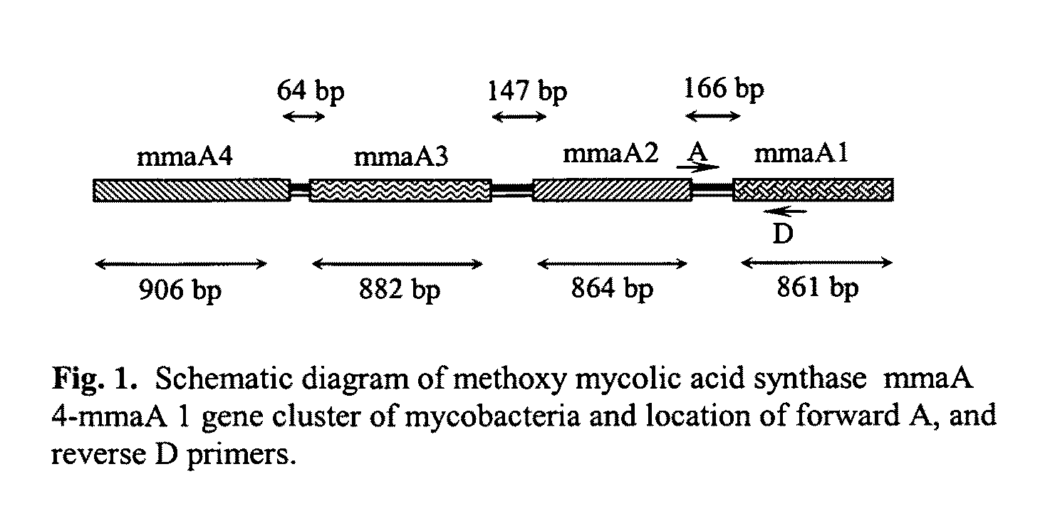
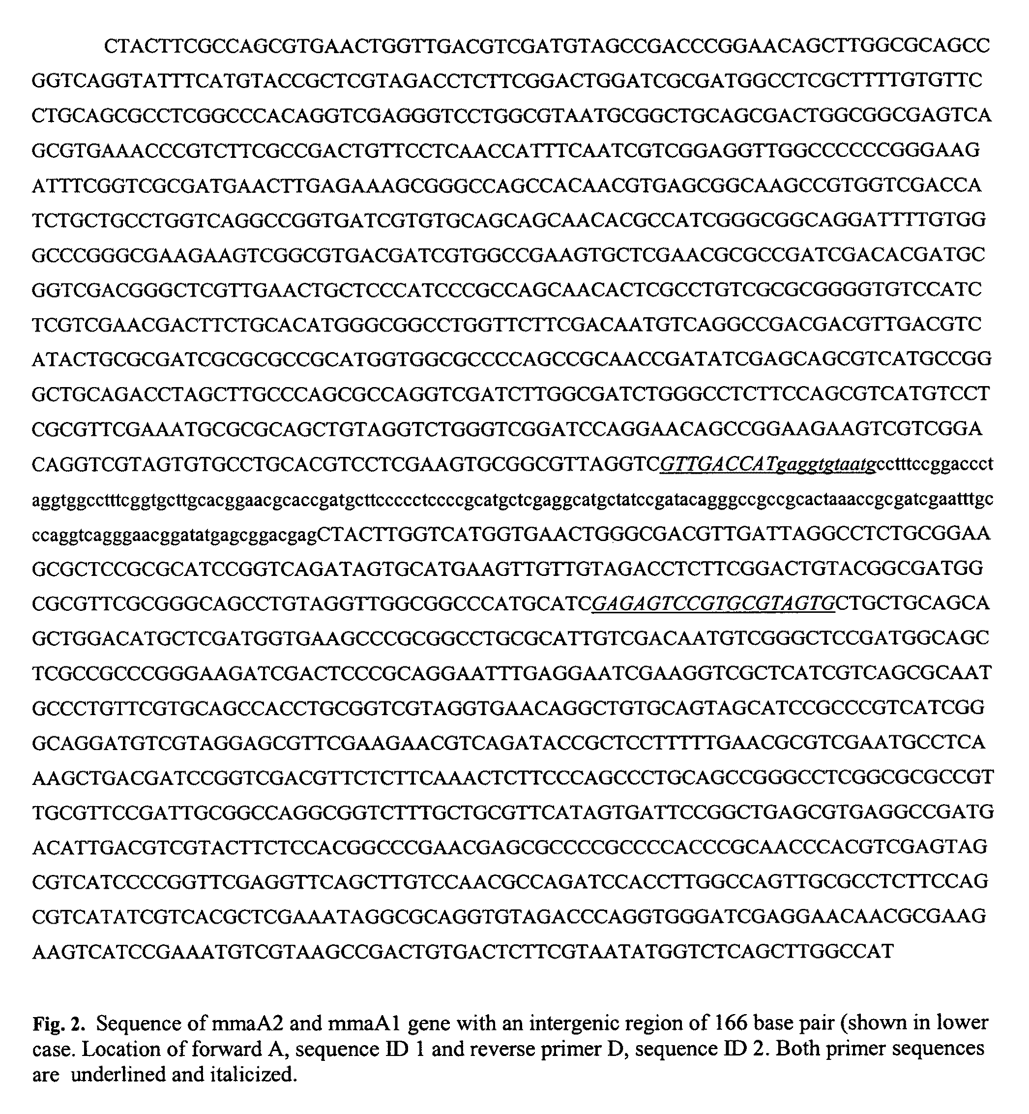
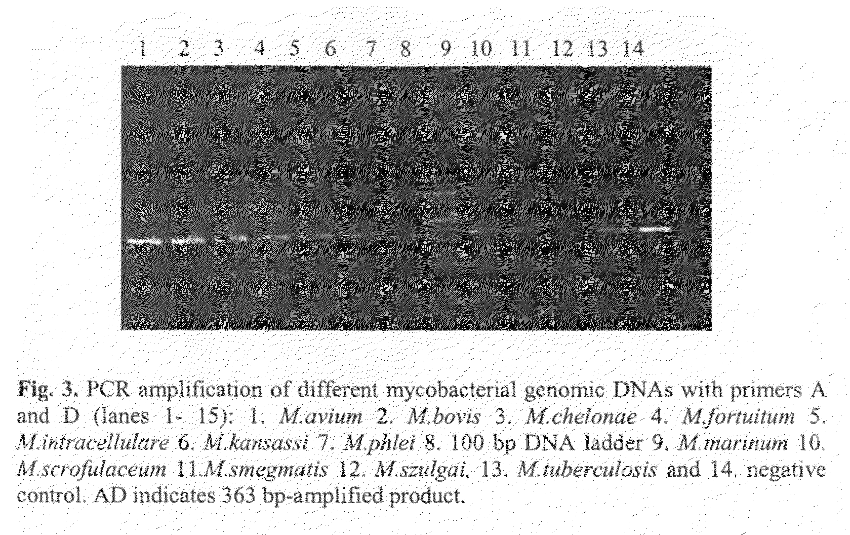
FIELD OF THE INVENTION[0001]The present invention relates to detection of pathogenic mycobacteria in clinical specimens such as sputum, cerebrospinal fluid, gastric lavage and tissue biopsies etc., wherein the novel stretch of DNA that lies in the intergenic region between methyl mycolic acid synthase genes mmaA1 and mmaA2 and the flanking region in mmaA1 and mmaA2 genes and is the invention uses a pair of designed oligonucleotide primers that specifically amplifies the target DNA from the clinical specimens.BACKGROUND OF THE INVENTION[0002]Tuberculosis is the number one killer disease. Every year it kills largest number of people due to a single infectious disease. According to a World Health Organization Report (WHO) over 8 million cases of tuberculosis are reported every year with over 2.9 million deaths[0003]Tuberculosis deaths were gradually declining till early 90's by the virtue of availability of potent anti-tubercular drugs. The tuberculosis cases and deaths are again on ri...

Claims


Application Information

Patent Timeline
Patent Type & Authority Patents(United States)
IPC IPC(8): C12P19/34C07H21/04C12Q1/00C12Q1/68
CPCC12Q1/689C07H21/04
Inventor DAS, RAKHA HARIKUMARSINGH, MEGHPATI
Owner COUNCIL OF SCI & IND RES
Learn More