Irradiation device and collimator

a collimator and irradiation device technology, applied in the field of irradiation device and collimator, can solve the problems of destroying surrounding tissue, affecting the health of the patient, damage to healthy tissue, etc., and achieves the effect of increasing the irradiation intensity, facilitating observation of tolerances, and increasing the pri

Active Publication Date: 2010-08-24
DEUTES KREBSFORSCHUNGSZENT STIFTUNG DES OFFENTLICHEN RECHTS
View PDF22 Cites 29 Cited by
  • Summary
  • Abstract
  • Description
  • Claims
  • Application Information

AI Technical Summary

Benefits of technology

[0042]This radiation application is preferably practiced by using single applications over a certain period with unchanged parameters. This bears the advantage that the application process is not disturbed by mechanical adjustment operations so that the required tolerances can be observed more easily.
[0043]The realization of application can be imagined in such a way that each single application captures a cylindrical area that extends through the whole human body. These cylindrical areas differ in size and come from all possible directions and intersect within the area of the object to be treated, so that this area will be exposed to radiation dose which is a multiple of that applied to the surrounding tissue. This corresponds to the image of filling a room with numerous cylinders of different size which are not only aligned differently within the space but occupy the space multiplicatively. Especially this combination of juxtaposed and overlaying positions and the combination of both in the area of the object to be treated produces the required increase in the irradiation intensity, whereby the surrounding tissue remains as unaffected as possible or will at least be able to regenerate, whereas the tissue to be treated, e.g. the tumor, will die off. Accordingly, it is also possible to avoid critical areas, such as nerves, at the margins of the object to be treated so that, if possible, no direct radiation will impinge on these regions.
[0044]One particularly useful aspect of the invention refers to the design of the iris diaphragms. In this context best contiguousness of the diaphragm leaves is achieved through the application of force that presses the side faces of the leaves together. This can be achieved, for example, if the diaphragm leaves consist of a shielding element made of shielding material and a bearing element, whereby the bearing element is provided with a guide element of a linear guiding mechanism for the adjustment movement, and the shielding element on the bearing element is supported with springs, so that it will be pressed evenly towards the direction of the adjacent diaphragm leaves. This structure has a great number of advantages: exact surface contact is ensured as the side faces always border each other evenly so that a possible gap can be caused only by an uneven or rough surface condition. Proper surface contact is guaranteed even in case of certain dimensional variations, so that no special requirements on production accuracy must be demanded which would lead to far higher prices. This makes the irradiation device considerably less expensive. Furthermore, jamming of the diaphragms and / or the adjusting mechanism can be excluded as far as possible, even if it comes to a certain deviation in tolerance. Also, it is improbable that this jamming will occur at a later point, for example as a result of wear, dirt, minor damage or thermal expansion. This not only reduces the costs of production, but also offers considerable improvement in the functional safety of the device.
[0045]The irradiation head that consists of the radiation source and the collimator, may be positioned on a gantry that enables irradiation of the object to be treated from different directions. Preferably, however, the irradiation head including the radiation source and the collimator, is mounted to a robot arm designed to maneuver the irradiation head into any position and angular alignment within a three-dimensional area. Compared to the previous arrangement of the irradiation heads on a gantry, the use of a robot arm offers the advantage of a considerably greater multitude of spatial positioning and alignment. Being mounted on a gantry, the irradiation head circulates around a center where the object to be treated is positioned. Further variations are possible only if the position of the patient is slightly changed, so that the focus can be brought to different positions on the object to be treated. In contrast, a robot arm does not need to be focused to a center, as it can be directed to any coordinates and spatial alignment or to any desired combination thereof, whereby the only restriction is the specific construction of the robot arm, which is, however, much less a limitation in positioning than that imposed by a gantry. Consequently, the robot arm offers a considerably higher degree of freedom in adjustment, so that optimum use can be made of the multiplex adjoining, overlaying and interlacing of partial spaces of different size as required by the single applications, and critical areas, such as nerves, can be better avoided.
[0046]To enable very quick and precise scanning of the beam position, the collimator may be installed on a track with a spherical surface, so that it can be shifted in all directions for pivoting motions within a limited spatial angle range, whereby the axis must always be oriented toward the radiation source. The installation of a collimator on such a guide with spherical surface has been published in DE 101 57 523 C1, which is hereby incorporated by reference. Certainly, this structure can be combined with a gantry or robot arm, so that control movements within a smaller range can be performed with this guide system and in a larger range with the gantry or the robot arm.
[0047]The design of the irradiation device may include that each iris diaphragm is equipped with a drive mechanism. Then, the design of the control unit may provide for adjusting the relevant aperture according to the divergence of rays. This may be useful, if the collimator with the iris diaphragms shall be supplied for installation in different types of irradiation devices so as to meet different distances to the radiation source. The control unit can be used to define how the apertures shall interact. In this way, the relevant differences in the divergence of rays can be considered by the program configuration. As an alternative, the iris diaphragms may be operated by means of one single drive mechanism. In this case, the control mechanism can adjust the relevant aperture according to the divergence of the rays. This requires less effort and possible programming errors can be excluded. This alternative is particularly useful, if the collimator with its iris diaphragms is permanently assigned to the same radiation source in an irradiation device.

Problems solved by technology

Since in most cases the tumors or other areas to be diagnosed are round in shape, the imaging field should be circular as well and, of course, it should not exceed the area of interest to the doctor, as X-rays are also harmful to the tissue.
Consequently, this radiation would also destroy the surrounding tissue.
Only high-energy therapeutic radiation—as required, for example, for the destruction of tumor tissue—will cause damage to healthy tissue in the over-range irradiation area, i.e. outside the tumor tissue to be irradiated, since radiation must be applied at a certain intensity and over a certain period of time required to destroy the tumor issue.
These over-range irradiation areas were caused by missing or insufficient imaging of the contour of the tumor tissue by the collimators and resulted from the occurrence of penumbrae at the borders of the area to be irradiated, since the shielding material was not in parallel alignment with the rays so that it could not provide full shielding, in particular when large areas were to be irradiated.
Furthermore, many irradiation devices showed leakage radiation emitting through the gap between adjacent shielding plates.
Moreover, large irradiation fields involved large penumbrae, since the block collimators were no longer in parallel alignment with the diverging optical path.
However, this type of collimator represents the shape of a tumor quite insufficiently and it does not offer a solution for the formation of penumbrae caused by the front edges of the diaphragm leaves which are not aligned in the direction of the optical path.
The prevention of leakage radiation is also insufficient.
Although the areas where the diaphragm leaves meet are provided with inset tongues, these tongues are too thin for an effective shielding of radiation emitting through the gap between adjacent leaves.
This type of reconstruction of the tumor shape or any other area to be irradiated is certainly quite inadequate and the penumbra problem also remains unsolved.
However, this collimator only allows formation of square-shaped irradiation fields, so that large exceeding radiation areas at the edges had to be accepted.
One disadvantage of this collimator is the intricate mechanical design required for the correct shifting and simultaneous turning of the plates in all planes so as to achieve a beam collimation aperture in the form of the frustum of a pyramid.
One further disadvantage of this solution is that the collimation field for irradiation can only be formed on the basis of polygons—depending on the number of plates—but the real tumor contour cannot be accurately defined.
The disadvantages included the complicated procedure with the continuous exchange of collimators, a prolonged use of expensive devices, as well as the costs for the production of a large number of collimators for each irradiation process; these collimators could not be used any further, since they were designed for one specific patient and could be used only within a very limited time frame even for this patient; the latter resulting from the continuous change of a patient's tumor by growth, regression or deformation.
At first sight, these multileaf collimators had the advantage of a quick setting for any type of shape, however, the mechanical design with the adjusting elements for each leaf was a considerable disadvantage, as well as the more or less large penumbra which occurred at each border of the irradiation field caused by a leaf, depending on its distance from the axis of the optical path.
This, however, requires an even more complicated mechanical structure of the multileaf collimator.
Although the collimator opening of the above proposal is small enough and prevents the formation of penumbrae, it requires extensive time for the scanning process—with a large diaphragm aperture the scanning process can be completed earlier, but the required accuracy cannot be achieved.
The use of multiple-hole plates to generate a beam of several scanning rays (FIGS. 5 and 5a) did not lead to satisfactory time reduction.
This leads either to a collimator of considerable weight or the number of aperture sizes must be limited, for example, to three openings.
The above stated reasons involve a further disadvantage of this collimator, since only a few defined collimator apertures are available so that the variability of beam collimation is quite limited.
In particular, the reduced number of openings makes it impossible to use large apertures of different diameters which could be used to treat one larger surface of the area to be irradiated first and then to irradiate the marginal areas with graded, smaller beams.
This is not only inconvenient for the patient who must be immobilized, but it also reduces the number of treatments possible with one device—an aspect of great economic significance in view of the high purchasing and operating costs of these devices.
Apart from this, the application accuracy in marginal areas is limited, which is critical if areas with nerves are in the vicinity.
This would cause a considerably increased complexity with regard to the calculation of the great number of individual irradiations to be applied—which often sum up to more than one hundred for one single irradiation session—and when considering the above-stated parameters aimed at achieving a specific three-dimensional irradiation profile.
But even if the different orientations of the polygonal beam shape were accepted without eliminating them, this would lead to errors in the irradiation profile applied.
These errors would increase with the deviation of the polygonal shape from a circular form.
As a consequence, however, integration in one diaphragm would require enormous mechanical efforts which, from a certain number of diaphragm leaves including drive and guide mechanisms for each leaf, would soon reach the limit of spatial accommodation.
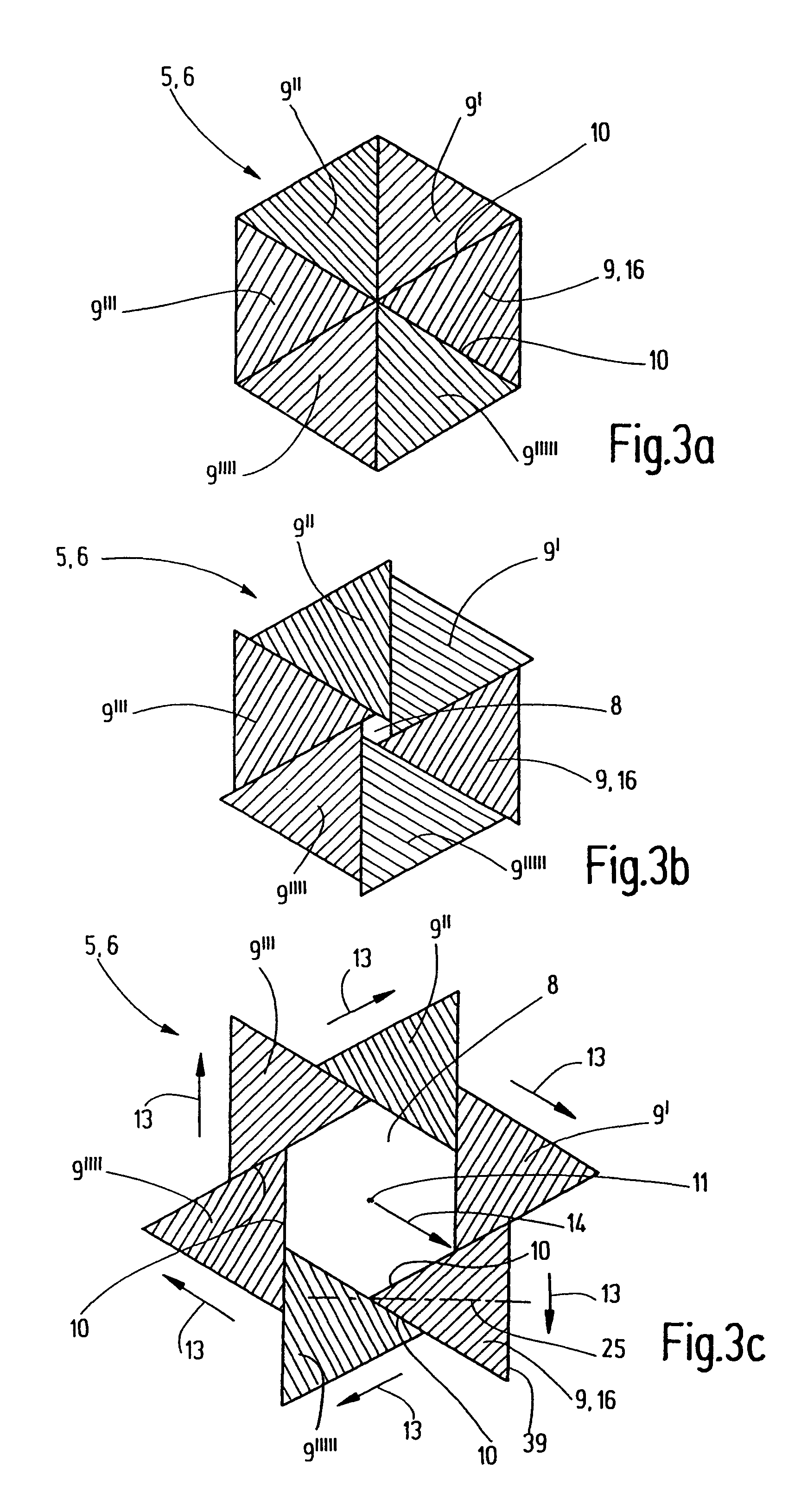
Since as many leaves as possible shall be used, the thickness of these leaves does not allow to put them in multiple layers one on top of the other.
Even if these side faces are intricately worked, a small gap will remain and cause an emitting of leakage radiation.
This could be prevented by tongues covering the gap as proposed in DE 20 53 089 A, but in view of the great number of required leaves this would increase complexity and require further installation space, since the tongues must be adequately thick so as to ensure adequate shielding properties.
However, this procedure would multiply the time required for processing and computation.
Apart from the use of an X-ray scanner for image generation, the most significant disadvantage of the irradiation device according to EP 0 382 560 A1 is, however, that it mentions a type of “scanning movement” by the therapy beam which is put into more concrete terms only insofar that an attempt is made to use the iris diaphragm to gain an approximate image of the object's shape by the form of the beam cross-section from each spatial angle.
Although this enables the formation of a round irradiation area, as well as the formation of irradiation regions with an elliptical cross-section (as shown in FIG. 4 of the above-mentioned publication), irregular regions, which the object to be treated usually are, cannot be achieved by a combination of individual applications in this manner.

Method used


Image

  • Irradiation device and collimator
  • Irradiation device and collimator
  • Irradiation device and collimator

Examples


Embodiment Construction

[0066]FIG. 1 shows a schematic illustration of the irradiation device, as defined by the invention, in a cross-sectional view. It shows the irradiation head 15 which includes a radiation source 3 and collimator 1 as defined by the invention. The drawing does not show the structure used to move the irradiation head 15 to different spatial positions. This structure may be a gantry, commonly-known in this field, which orientates the irradiation head 15 to an iso-center, so as to enable irradiation of an object to be treated 4, mostly a tumor, from all sides. When such a gantry is used, the focusing to the iso-center depends on its mechanical structure and variations are only possible by moving the patient, and thus the object to be treated 4. Instead of an orientation to one iso-center, the invention recommends a design variant with the irradiation head 15 being mounted on a robot arm that enables operation to all spatial coordinates and in any spatial angle alignment as far as is allo...


PUM


Abstract

An irradiation device for radiation treatment and a collimator (1) are used to define a beam of high-energy rays (2) with beam limitation by means of an iris diaphragm (5) having adjusting elements (7), and a mechanism that is used to direct the beams (2′) limited by the collimator (1) to the object to be treated (4) from all sides, wherein the parameters for direction, surface area, intensity and time of irradiation can be specified with the help of a control mechanism. An exact three-dimensional irradiation profile is produced, while keeping the costs and efforts regarding mechanical engineering, computation and irradiation time low. This is achieved by using at least one further iris diaphragm (6) located in coaxial alignment in the optical path, whereby the leaves (9, 9′, 9″, 9′″, 9″″, 9′″″) of the iris diaphragms (5, 6) are arranged in a staggered manner and in a rotational sense around their axis (11), so that the beam limited by the collimator (1) has the cross-section of a polygon (12), the number of corners of which complies with the number of the leaves of all iris diaphragms (5, 6), so that the leaves (9, 9′, 9″, 9′″, 9″″, 9′″″) allow for linear adjustment movements (13), the control mechanism being suitable to enable the irradiation of an irregular space through the overlaying and adjoining of many irradiated spaces (28).

Description

[0001]This application is the national stage of PCT / EP2006 / 007471 filed on Jul. 27, 2006.BACKGROUND OF THE INVENTION[0002]The invention relates to an irradiation device with a collimator used to define a beam of high-energy rays proceeding from an essentially punctiform radiation source and directed to an object to be treated, and which serves for radiation treatment, in particular the stereotactic conformation radiotherapy of tumors, whereby the collimator is realized by means of iris diaphragms with adjusting elements so as to provide for variable apertures for beam collimation, and a mechanism that shall be used to direct the beams limited by the collimator to the object to be treated from all sides, and whereby the parameters for direction, surface area, intensity and time of irradiation can be controlled in such a manner that a three-dimensional dosing profile for radiation application can be achieved.[0003]Furthermore, the invention relates to a collimator used to define a bea...

Claims


Application Information

Patent Timeline
Patent Type & Authority Patents(United States)
IPC IPC(8): A61N5/10G21K1/04
CPCA61N5/1042G21K1/04
Inventor ECHNER, GERNOT
Owner DEUTES KREBSFORSCHUNGSZENT STIFTUNG DES OFFENTLICHEN RECHTS
Why Eureka
Try Eureka