Optical detection of intravenous infiltration

a technology of intravenous infiltration and optical detection, which is applied in the field of optical detection of intravenous infiltration, can solve the problems of tissue necrosis, skin grafting, or amputation, necrosis of skin debridement, and precipitation of significant scarring around joints, so as to achieve less complex and more reliable results

Active Publication Date: 2010-11-02
IVWATCH
View PDF7 Cites 95 Cited by
  • Summary
  • Abstract
  • Description
  • Claims
  • Application Information

AI Technical Summary

Benefits of technology

[0019](6) That uses a single light source and a single detector making the

Problems solved by technology

Severe infiltration may lead to necrosis requiring skin debridement, skin grafting, or amputation.
The leakage of cytotoxic drugs, intravenous nutrition, solutions of calcium, potassium, and bicarbonate, and even 10% dextrose outside the vein into which they are delivered is known to cause tissue necrosis and to precipitate significant scarring around joints.
Because the amount of connective tissue is limited in elderly patients as well as the very young, they are prone to extensive diffusion of infiltrated fluid.
One study reported that infiltration results in skin loss in 0.24% of the peripheral lines.
The visual and tactile examination technique is ineffective in detecting infiltration, since by the time infiltration is det

Method used


Image

  • Optical detection of intravenous infiltration
  • Optical detection of intravenous infiltration
  • Optical detection of intravenous infiltration

Examples


Embodiment Construction

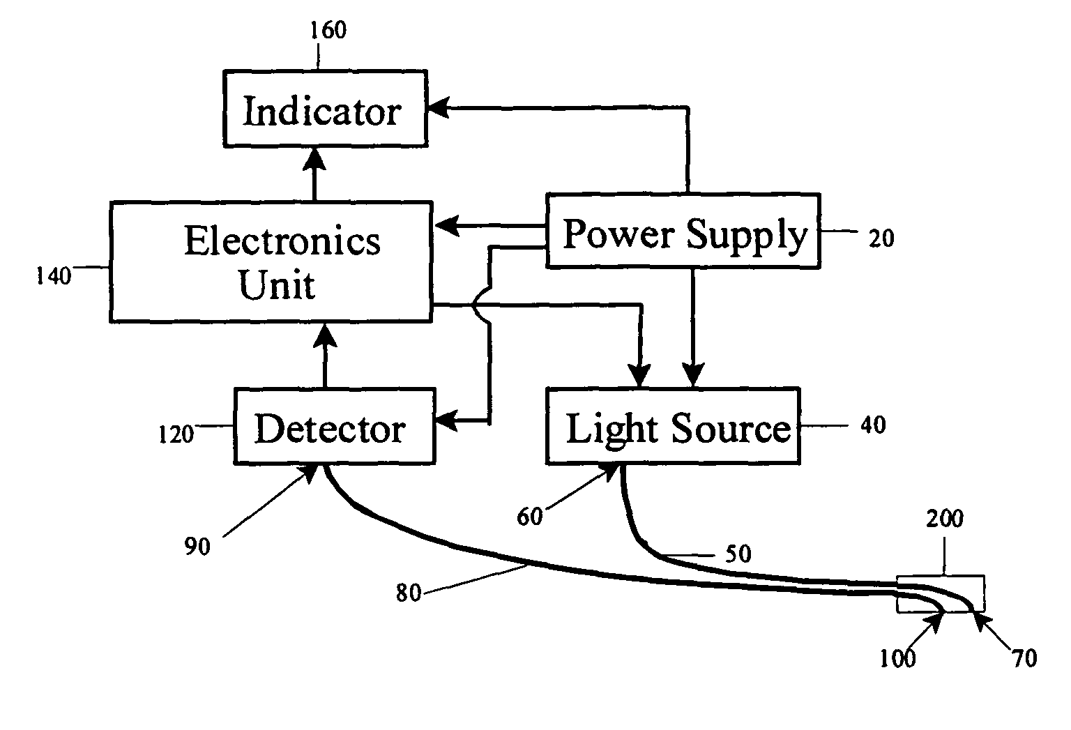
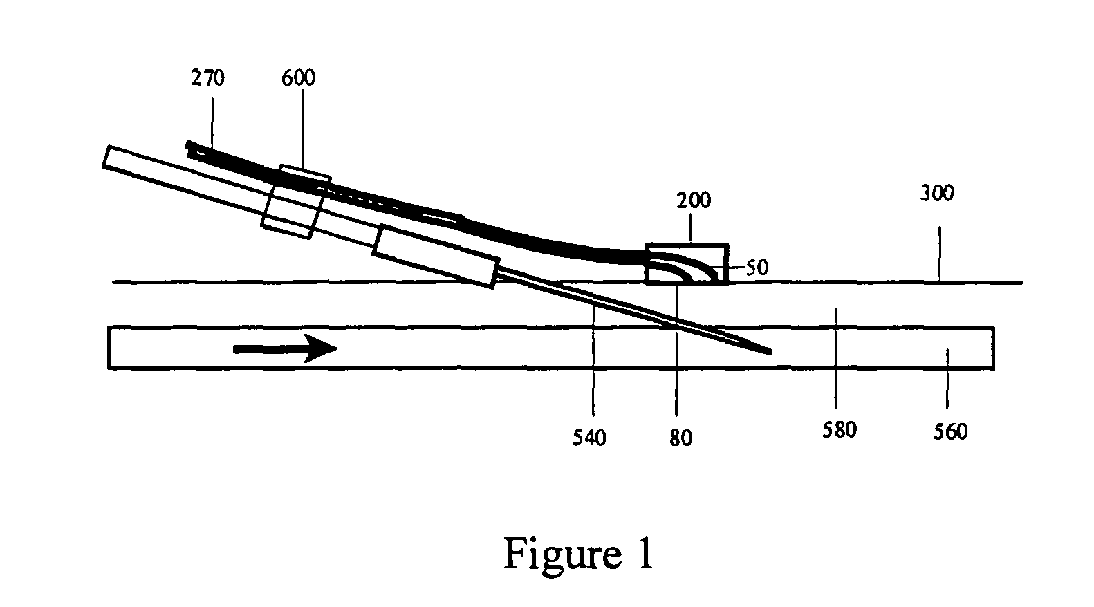
[0033]As shown in FIG. 1, when a beam of optical radiation impinges on skin 300 near an IV infusion site, the radiation reflected, scattered, diffused or otherwise emitted from the skin can be measured. As shown in the figure, an optical fiber bundle 270 comprises an illumination light guide 50 that provides illumination to the infusion site and a collection light guide 80 that collects the electromagnetic radiation reflected from the infusion site. The ends of the light guides 50 and 80 are embedded in a skin-contact sensor 200 that is secured onto the skin 300. A needle 540 is inserted through the skin 300 into a vein 560 for infusion of IV fluids.

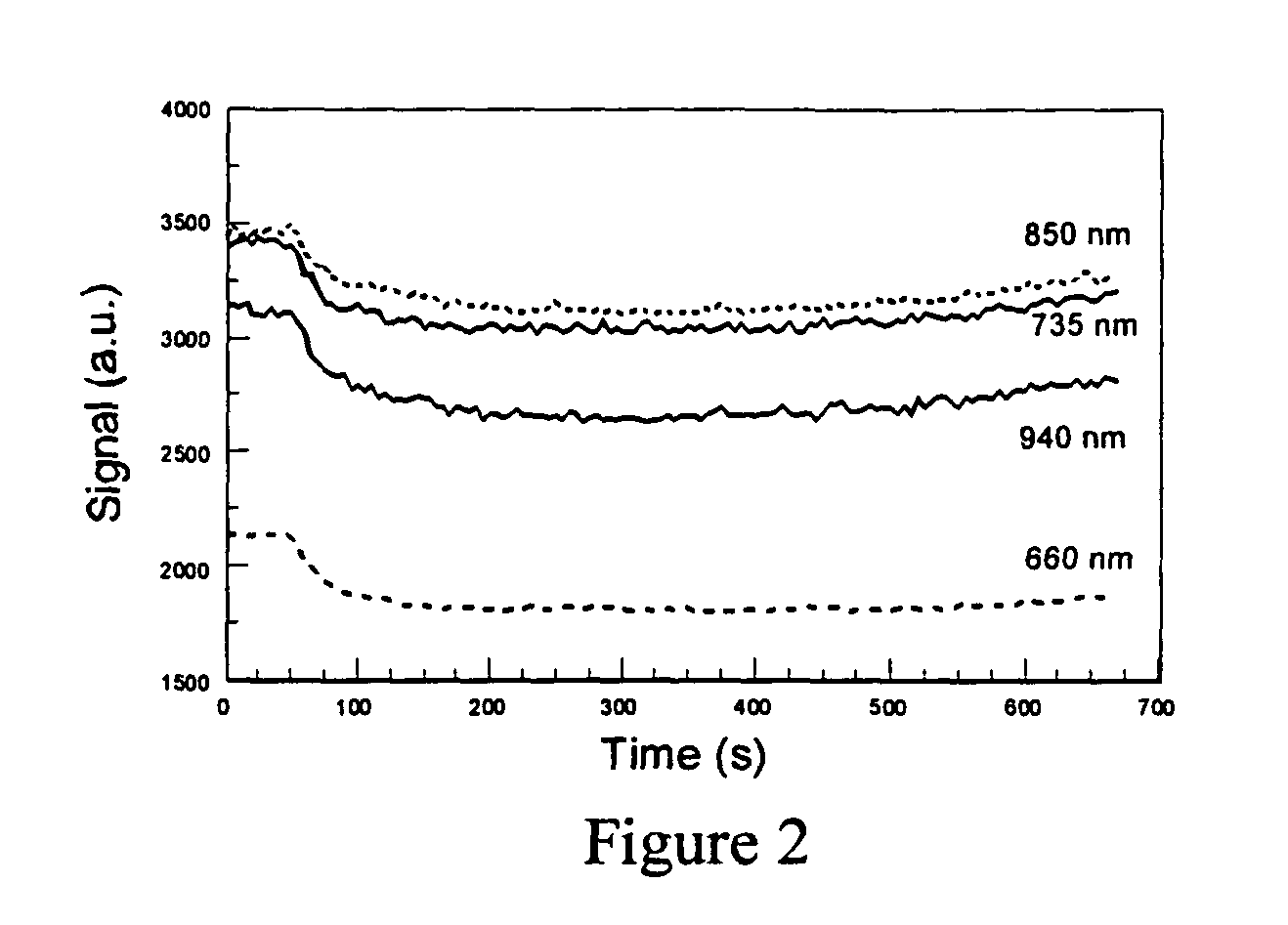
[0034]When IV fluid infiltrates the interstitial tissue space, optical density of tissue changes. This change can be measured as follows. First, the infusion site is illuminated using a beam of electromagnetic radiation with certain wavelength. Before energizing the illumination source, the radiation collected after insertion of the need...


PUM


Abstract

An intravenous infiltration detection apparatus for monitoring intravenous failures, which applies an optical method coupled with fiber optics and algorithms for tissue optics to provide a means for noninvasive detection of intravenous infiltration surround the site of IV injection. In the invention, the tissue surrounding the injection site is exposed to a single-wavelength of electromagnetic radiation, and light is collected with only one detector. Changes in the relative intensity of the radiation reflected, scattered, diffused or otherwise emitted provide a means for monitoring infiltration. The invention provides routine, automated, continuous, and real-time monitoring for patients undergoing IV therapy.

Description

BACKGROUND OF THE INVENTION[0001]In the United States, approximately 80% of hospital patients require intravenous (IV) therapy and approximately 50% of the IV lines fail due to infiltration, a clot in the cannula, an inflammatory response of the vein, or separation of the cannula from the vein. IV infiltration is usually accompanied by pain, erythema, and / or swelling at the cannula tip or the insertion site. Severe infiltration may lead to necrosis requiring skin debridement, skin grafting, or amputation. One common area of malpractice lawsuits filed against physicians and nurses involves infiltration. The leakage of cytotoxic drugs, intravenous nutrition, solutions of calcium, potassium, and bicarbonate, and even 10% dextrose outside the vein into which they are delivered is known to cause tissue necrosis and to precipitate significant scarring around joints. An infiltration rate of 0.1-1% has been reported in cases where contrast agents were used in medical imaging procedures. Ear...

Claims


Application Information

Patent Timeline
IPC IPC(8): A61B6/00A61B5/05
CPCA61B5/0059A61M5/158A61M5/16836A61B2560/0276A61M2005/1588A61M2205/3313
Inventor WINCHESTER, JR., LEONARD W.CHOU, NEE-YIN
Owner IVWATCH
Why Eureka
Try Eureka