Receptor for GDNF family ligands

a family ligand and receptor technology, applied in the field of new receptors for gdnf family ligands, can solve the problems that other studies have failed to show strong clinical benefits for gdnf, and achieve the effect of preventing or delaying a neurodegenerative process and better biodistribution of gfls

Inactive Publication Date: 2011-10-11
SAARMA MART +3
View PDF1 Cites 3 Cited by
  • Summary
  • Abstract
  • Description
  • Claims
  • Application Information

AI Technical Summary

Benefits of technology

[0008]The present invention also provides a complex for identifying a compound, which interferes with the interaction between a GFL and a HSPG, wherein said complex comprises at least one immobilized GFL and at least one heparan sulfate proteoglycan (HSPG) anchored to a matrix. The complex can be used in neuronal tissue or in neuronal cells. As anchored GFL-HSPG complexes GFLs are useful in for identifying molecules which interfere with the interaction between a GFL and a HSPG, preventing the binding of GFLs to HSPG leading to a better biodistribution of the GFLs, GDNF, neurturin and artemin. The anchored complex can be attached to a solid support.
[0009]The present invention also provides a method for preventing or delaying a neurodegenerative process, wherein a molecule, which is identified with the method of claim 1 and interferes with the interaction between a glial cell-line derived neurotrophic factor family ligand (GFL) and a HSPG, is administered to a subject in need, in an amount, which is capable of replacing said GFL forming a complex between at least one immobilized GFL and at least one heparan sulfate proteoglycan (HSPG), whereby leading to release of GFL and increased biodistribution of GFL. In a preferred embodiment the neuronal cells are cortical neurons, more preferably embryonic cortical neurons. In another embodiment the matrix anchored complex between immobilized GFL and heparan sulfate proteoglycan (HSPG) comprises syndecan-3 or oligomerized syndecan-3. In one embodiment the neuronal cell is a cortical neuron, more preferably an embryonic cortical neuron. In another embodiment the matrix anchored complex between immobilized GFL and heparan sulfate proteoglycan (HSPG) comprises syndecan-3 or oligomerized syndecan-3.

Problems solved by technology

Even if GFLs, particularly GDNF, have shown promising results in treatment of Parkinson's disease (Gill et al., 2003) and amyotrophic lateral sclerosis (ALS) (Henderson et al., 1994), other studies have failed to show strong clinical benefits for GDNF (Lang et al., 2006).

Method used


Image

  • Receptor for GDNF family ligands
  • Receptor for GDNF family ligands
  • Receptor for GDNF family ligands

Examples


example 1

Cell Cultures and Reagents

[0084]Rat glioma C6 cell line and mouse neuroblastoma cell line Neuro2a were from ATCC. Early passage RET-deficient human SHEP cells were provided by Dr. Marc Billaud (CNRS, Lyon, France). MG87-RET cells were provided by Dr. Carlos F. Ibáñez (Karolinska Institute, Stockholm, Sweden). Rat hippocampal neurons were isolated from E17 embryos. Mouse cortical neurons were isolated from E15 embryos. Low molecular weight heparin, heparin-BSA, EDAC / NHS crosslinker, lactoperoxidase, PI-PLC, chondroitinase ABC, globulin-free BSA and cell culture tested BSA were purchased from Sigma. SU6656 was bought from Calbiochem. Heparinase III was from Seikagaku Corp. (Seikagaku's trademark for this product is heparitinase I). The dominant-negative Src adenoviral construct was a kind gift of Dr. David Kaplan (Hospital of Sick Child, Toronto, Canada). Desulfated heparins were kindly provided by Dr. Ryo Takano (Okinawa National College of Technology, Nago, Okinawa, Japan).

example 2

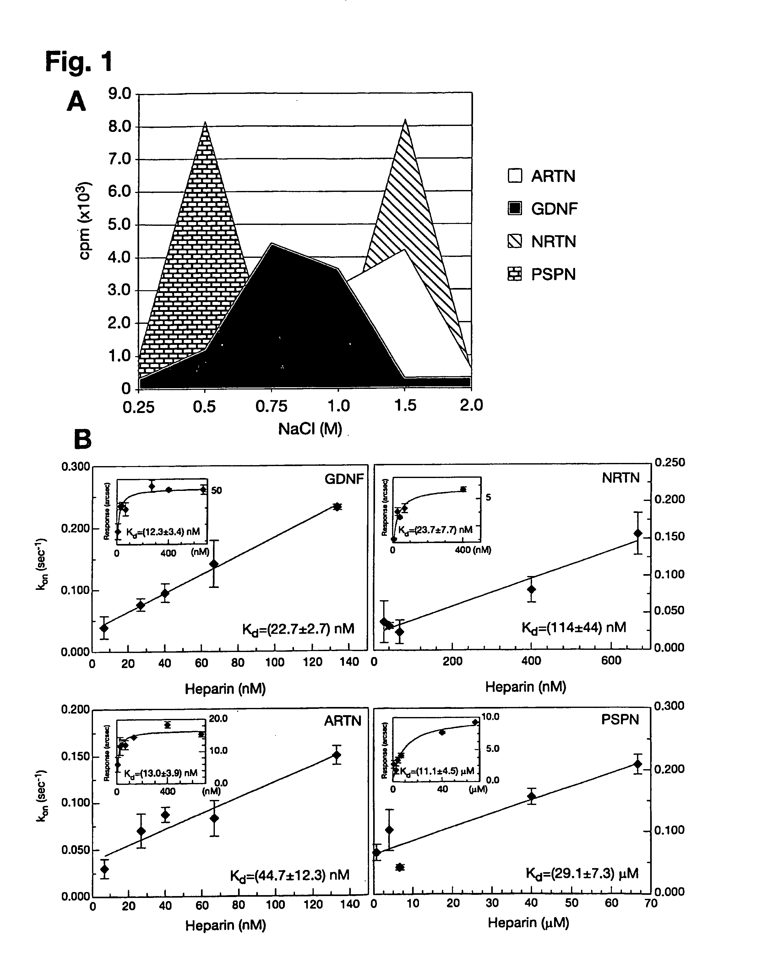
GFL Iodination and Binding to Heparin-Sepharose

[0085]GFL iodination was performed as described (Lindahl et al., 2001). Heparin-sepharose column (Amersham Biosciences) chromatography was performed to assess GFL binding to heparin. GDNF, ARTN, NRTN and PSPN were purchased from PeproTech, Ltd. or R&D Systems, Inc. GFLs were enzymatically labeled with carrier-free [125I]NaI (Amersham Biosciences) by the lactoperoxidase method to a specific activity of 100000-200000 cpm / ng as described elsewhere (Lindahl et al., 2001). 15000-20000 cpm of iodinated GFLs, together with 10 μg of the same unlabeled protein as a carrier, were injected into a 1 ml heparin-sepharose column (Amersham Biosciences) in phosphate buffer pH 7.2 containing 100 mM NaCl and 10 mg / ml of BSA. After washing with 5 volumes of the same buffer, iodinated GFLs were eluted from the column by step elution (2 ml) with increasing NaCl concentration. The resulting fractions were counted on Rackbeta 1214 (LKB / Wallac) scintillation c...

example 3

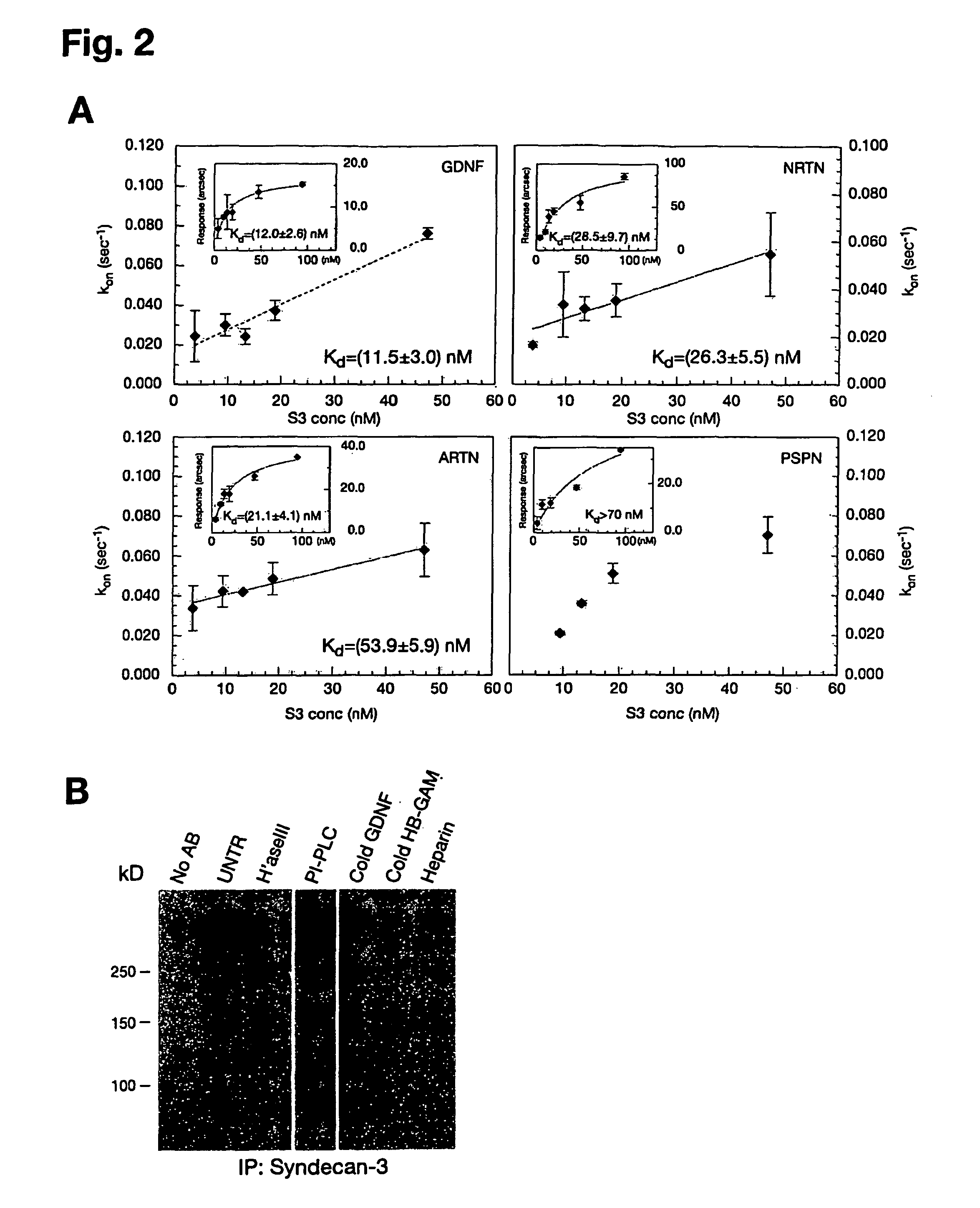
Surface Plasmon Resonance (SPR) Studies

[0086]The affinities of GFLs for heparin and heparin-like molecules (e.g. syndecan-3) were determined by plasmon surface resonance using the IAsys system (Affinity Sensors, Cambridge, UK). Planar aminosilane cuvettes (Affinity Sensors) were used for protein coupling to avoid problems with mass transport, which occur frequently with matrix surfaces. In the initial approach, heparin-BSA was immobilized to the cuvette surface using the manufacturer's instructions and different ligand concentrations were used to determine association (ka) and dissociation (kdis) rates using the IAsys Fastfit software. The dissociation constant Kd was determined from the binding dynamics as Kd=ka / kdis, or from equilibrium response (R) using a simple Langmuir fit: R=(Rmax*C) / (Kd+C), where C is the ligand concentration. Because all ligands except PSPN displayed background binding to a control BSA cuvette, Kds were also determined in a system where GDNF, ARTN, NRTN and...


PUM

PropertyMeasurementUnit
concentrationaaaaaaaaaa
pHaaaaaaaaaa
dissociation constantsaaaaaaaaaa

Abstract

The present invention relates to an in vitro method for identifying a molecule, which interferes with the interaction between a glial cell-line derived neurotrophic factor family ligand (GFL) and a heparan sulfate proteoglycan (HSPG), by screening a library of molecules against a matrix anchored complex comprising at least one immobilized glial cell-line derived neurotrophic factor family ligand (GFL) and at least one heparan sulfate proteoglycan (HSPG), wherein the interfering molecule is isolated based on its capacity to replace a glial cell-line derived neurotrophic factor family ligand (GFL) in said anchored complex. The invention also relates to a complex for identifying such a molecule. The invention also relates to methods for preventing or delaying a neurodegenerative process as well as to method for prophylactic treatment or treatment of a disorder in the nervous system.

Description

FIELD OF THE INVENTION[0001]The present invention is generally directed to a novel receptor for GDNF family ligands (GFLs). It provides methods for identifying a molecule, which interferes with the interaction between a glial cell-line derived neurotrophic factor (GDNF) family ligand (GFL) and a heparan sulfate proteoglycan (HSPG) in order to increase the biodistribution of GFLs. The present invention further provides a complex for identifying said molecule. The present invention further provides a method for preventing or delaying a neurodegenerative process as well as a method for prophylactic treatment or treatment of a disorder in the nervous system by administering the molecules of the present invention. The present invention further provides a method for identifying a molecule, which activates heparan sulfate proteoglycan (HSPG) and triggers intracellular signaling of the Src family kinase (SFK) signaling pathway, whereby leading to spread and migration of neuronal cells and n...

Claims


Application Information

Patent Timeline
Patent Type & Authority Patents(United States)
IPC IPC(8): G01N33/68
CPCG01N33/5308G01N33/543G01N2333/4722G01N2500/02G01N2800/28
Inventor SAARMA, MARTRAUVALA, HEIKKIBESPALOV, MAXIMTUMOVA, SARKA
Owner SAARMA MART
Why Eureka
Try Eureka