Method of gene transfer via vascular system or ureter

a technology of ureter and vascular system, applied in the direction of prosthesis, peptide/protein ingredients, immunological disorders, etc., can solve the problems of prolonged ischemia, chronic rejection, and shortened ischemia, and achieve the effect of reducing adverse effects and advantageous in cos

Inactive Publication Date: 2012-09-11
CHUGAI PHARMA CO LTD
View PDF16 Cites 5 Cited by
  • Summary
  • Abstract
  • Description
  • Claims
  • Application Information

AI Technical Summary

Benefits of technology

[0008]The present invention relates to a method for ex vivo gene transfer through a vascular system or a ureter. In a particularly preferable embodiment, the present invention provides a method for introducing and expressing, by electroporation, a desired gene in the whole organ used for organ transplantation. In particular, an ex vivo HGF gene therapy that utilizes electroporation aiming to protect the transplanted kidney is provided. According to the ex vivo HGF gene therapy method utilizing electroporation of the present invention, the gene is introduced only into the transplanted kidney. Namely, the method enables continuous reaction of HGF on the targeted local site, the kidney. Thus, the method is effective in reducing adverse effects compared to systemic administrations such as intravenous injection. In addition, the method of the present invention is safe because it does not require the use of a virus vector, and advantageous in terms of cost compared to administrations of protein drugs.
[0009]Because of intensive studies to resolve the above issues, the present inventors have succeeded in introducing a desired gene into an extirpated kidney by filling the renal artery and / or vein, or the ureter of the kidney with a solution containing the gene. Specifically, the inventors revealed that filling the renal artery and / or vein, or the ureter of the extirpated kidney with a solution containing a desired gene, and conducting electroporation on the whole kidney result in introduction of the gene into all glomerulus and interstitial cells with high efficiency. Thus, according to the present method a desired gene is safely and effectively introduced evenly into the glomerulus and interstitial cells.
[0010]Rejection of a transplanted kidney is effectively suppressed by introducing a gene encoding a protein having organ damage suppressing function, such as HGF gene, into an extirpated kidney and subsequently transplanting this kidney that had received gene transfer according to the present method. The ex vivo gene transfer method of the present invention has several advantages over in vivo gene transfer wherein a gene is directly introduced into the kidney. One advantage of the present method is that it enables ex vivo introduction of a gene after removing a kidney for transplantation that prevents gene transfer in to other organs. As a second advantage, the influence of electric shock on humans (burn or muscle contraction) caused by the electric pulses used for electroporation can be avoided. The third advantage is that the procedure can be performed during transplantation, and thus no complicated manipulations, such as puncture into renal artery and insertion of cannula, are required. In addition, transplantation that uses the method of the present invention is applicable for xenogeneic transplantation of pig kidneys to humans.
[0036]In particular, the present invention revealed that nucleic acids injected through the renal artery is specifically introduced into mesangial cells of the kidney. Thus, the present invention provides a method for introducing a desired gene into mesangial cells, comprising the steps of injecting an aqueous solution containing an expression vector comprising the desired gene into the renal artery, and introducing the expression vector into an injection area. The use of electroporation allows efficient introduction of the expression vector.
[0041]The method for gene transfer of the present invention particularly shows an excellent effect to continuously express a gene for a long period following the introduction of the gene into an organ. The present invention provides a method for expressing a desired gene in an organ for a long term, which method comprises the steps of injecting an aqueous solution containing an expression vector comprising the desired gene into the vascular system or a ureter of the target organ, and introducing the expression vector into an injection area. According to the method, the gene is introduced through the injected vascular system or the ureter into, for example, an area in proximity to a blood vessel, an interstitial region and / or a surrounding area of epithelial cells. The expression vector may be preferably introduced into the injection area by electroporation. Electroporation may be performed, for example, using forceps-type electrodes or a bathtub-type electrode. The method of the present invention is particularly suitable for ex vivo gene transfer, in which a vector is preferably introduced into an organ by electroporation using a bathtub-type electrode. Specifically, electroporation may be performed under the conditions described above. The target organ for gene transfer is not limited similarly as described above, and includes kidney, liver, spleen, heart and lung, but kidney is particularly preferable. The organ may be derived from any desired mammal. However, as non-human organs for transplantation into humans, pig organs are preferable. The pig organs used in the present invention may include those that have been genetically engineered to suppress the immune reaction that results upon transplantation into humans. The method of the present invention for long-term expression accomplishes an expression of the introduced gene for 3 days or longer, preferably 1 week (7 days) or longer, more preferably 2 weeks (14 days) or longer, more preferably 1 month (30 days) or longer, more preferably 2 months (60 days) or longer, and even more preferably 3 months (90 days) or longer. A vector incorporating viral latent infection machinery or a strong expression vector is suitable as the expression vector of the present invention. Specifically, preferable latent infection machineries include those of EB virus, i.e., EBNA-1 gene or oriP sequence, or combination thereof. Preferable strong expression vectors include expression vectors carrying the cytomegalovirus enhancer and chicken β-actin promoter. The gene to be introduced is not limited, but introduction of a gene encoding a protein having organ damage suppressing function enables to suppress tissue damages of an organ for a long term. Therefore, such a method enables to protect the transplanted organs from cytotoxicity and achieves long-term survival following organ transplantation.

Problems solved by technology

Currently, most important issue in kidney transplantation is abolition of renal function (renal death) due to chronic rejection.
This results in prolonged ischemia, leading to chronic rejection and poor long-term prognosis of renal functions.
However, HGF protein has a short half-life in blood.
Although continuous injection of protein drugs has merit in that a constant concentration can be maintained in blood, it requires the attachment of a catheter for a long term, which involves results in complex manipulation and risk of infection as well as restraint on patient's movement.
However, the safety issue remains to be solved for these methods due to their use of virus vectors for gene transfer.
However, this method has certain disadvantages, such as tissue damage.
In addition, gene expression is limited to the injected site and thus it is impossible to express the desired gene in the whole of the transplanted kidney.
However, the introduction of an HGF gene into a transplanted kidney to examine the survival rate has not been reported nor is there any report of the effectiveness of ex vivo gene transfer via electroporation.

Method used


Image

  • Method of gene transfer via vascular system or ureter
  • Method of gene transfer via vascular system or ureter
  • Method of gene transfer via vascular system or ureter

Examples


example 1

Introduction of FITC-Labeled Oligo DNA into Rat Kidney (1)

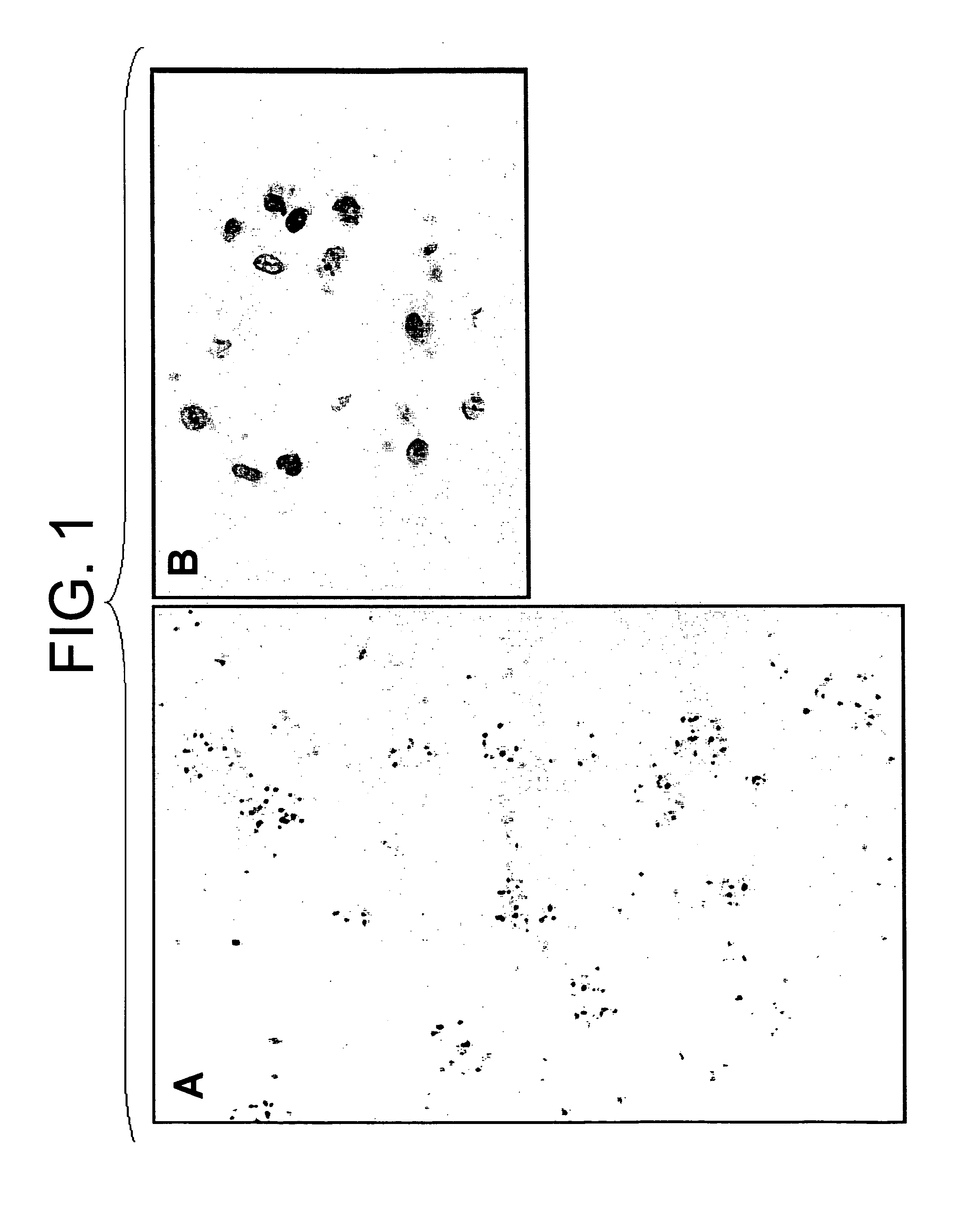
[0065]A twelve base pair oligo DNA fluorescently labeled with FITC at the 5′-terminus (5′-FITC-CGAGGGCGGCATGGG-3′; SEQ ID NO: 1) was dissolved in buffer (137 mM NaCl, 5.4 mM KCl, 10 mM Tris-HCl [pH 7.6]; hereinafter abbreviated as “BSS”) at a concentration of 50 μg / 500 μl for gene transfer. Sprague Dawley (hereinafter abbreviated as “SD”) rat (6-week-old male) was anesthetized by intraperitoneal administration of pentobarbital (50 mg / kg). After dissection of the abdomen, a kidney was exposed and a 24 G catheter (Terumo Corporation) was inserted into the renal artery for perfusion of the kidney with physiological saline and the oligo DNA described above was injected. Immediately after the injection of the oligo DNA, the renal vein was ligated with a clip. The kidney introduced with the DNA was nipped with the forceps-type electrodes to conduct gene transfer by electroporation applying a square-shaped voltage (75 V, 100 millise...

example 2

Introduction of FITC-Labeled Oligo DNA into Rat Kidney (2)

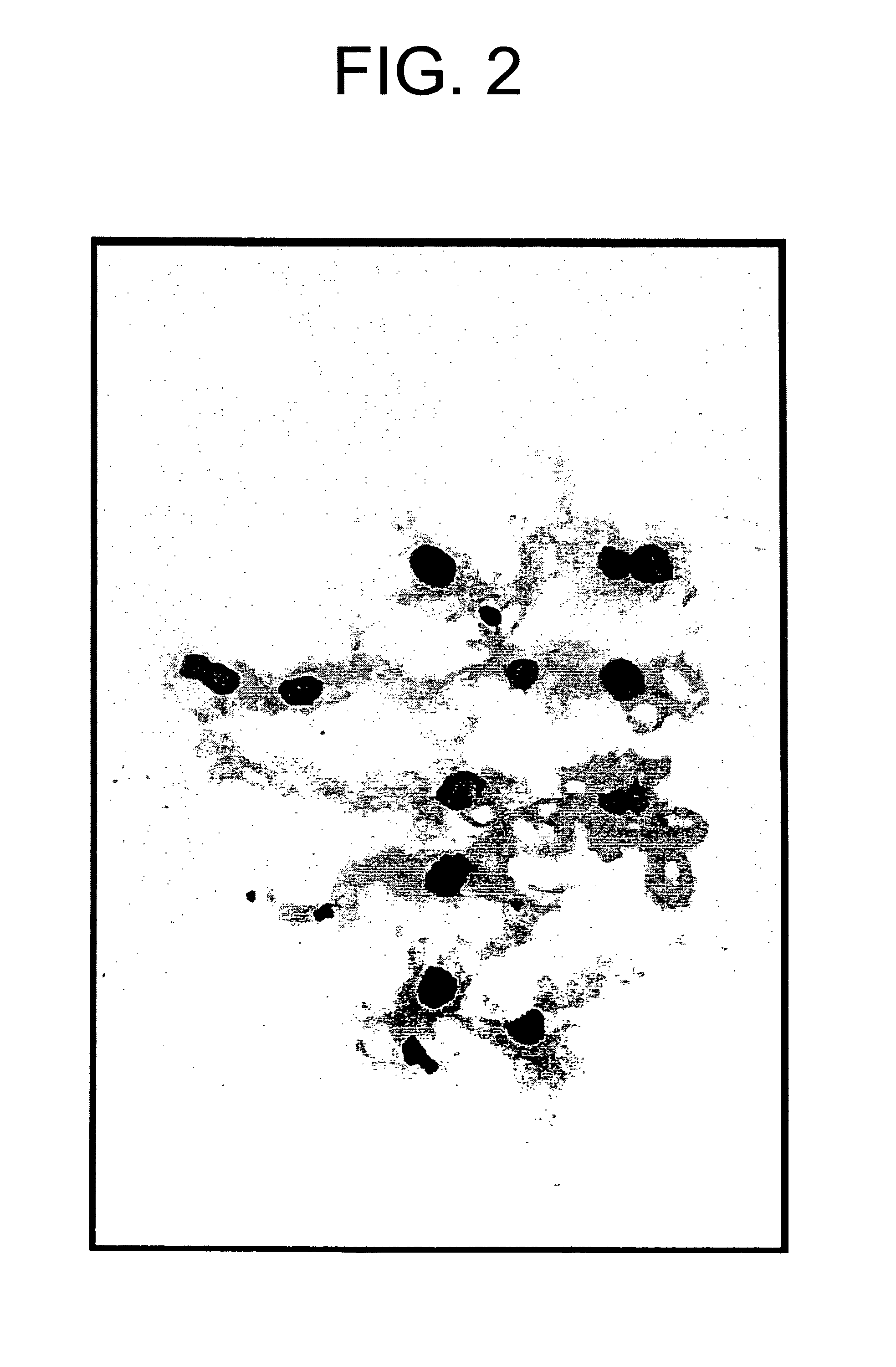
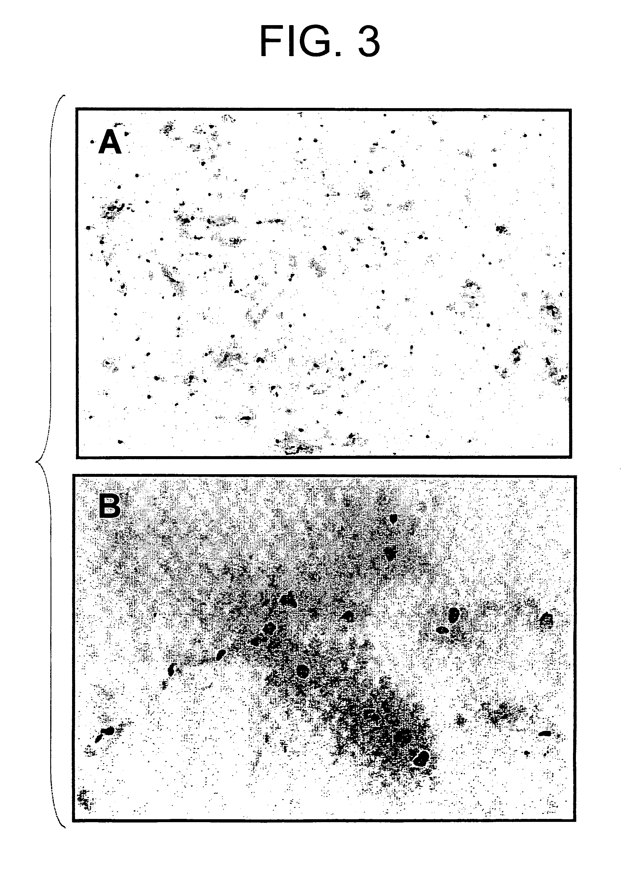
[0066]A twelve base pair oligo DNA fluorescently labeled with FITC at the 5′-terminus (5′-FITC-CGAGGGCGGCATGGG-3′; SEQ ID NO: 1) was dissolved in BSS at a concentration of 50 μg / 500 μl for gene transfer. SD rat (6-week-old male) was anesthetized by intraperitoneal administration of pentobarbital (50 mg / kg). After abdominal dissection, the left kidney and left ureter were exposed, a 30 G needle (Terumo Corporation) was inserted into the ureter, and the renal artery and vein were clamped with clips to inject the above-described oligo DNA into the kidney. The kidney introduced with the DNA was nipped with forceps-type electrodes to perform gene transfer by electroporation applying a square-shaped voltage (75 V, 100 milliseconds) six times with intervals of 900 milliseconds. Ten minutes after the gene transfer, the kidney was removed and observed under a fluorescent microscope. As shown in FIG. 3, the FITC-labeled oligo DNA was i...

example 3

Introduction of Luciferase Gene into Rat Kidney (1)

[0067]An expression vector carrying a latent infection machinery of EB virus inserted with a luciferase gene was constructed. The expression vector was dissolved in BSS at a concentration of 200 μg / 500 μl for gene transfer. SD rats (6-week-old males) were anesthetized by intraperitoneal administration of pentobarbital (50 mg / kg). After abdominal dissection, kidneys were exposed and 24 G catheter was inserted into the renal artery for perfusion of the kidneys with physiological saline and the above-described expression vector was injected. Immediately after the injection of the expression vector, the renal vein was ligated with a clip. The kidneys introduced with the gene were nipped with forceps-type electrodes to perform gene transfer by electroporation applying a square-shaped voltage (25, 50, 75 or 100 V, 100 milliseconds) six times with intervals of 900 milliseconds. The catheter was removed after the gene transfer, and the punc...


PUM

PropertyMeasurementUnit
voltageaaaaaaaaaa
voltageaaaaaaaaaa
voltageaaaaaaaaaa

Abstract

The present invention provides a method for gene transfer through the vascular system or a ureter. In a preferable embodiment of this invention, an aqueous solution containing an expression vector comprising a desired gene is injected through the vascular system or a ureter of a desired organ to introduce the gene into the injected area. Specifically, the method of the present invention is effective for ex vivo gene therapy wherein the hepatocyte growth factor (HGF) gene is introduced by electroporation into a kidney to be transplanted. According to the present invention, the HGF functions continuously on the kidney to be transplanted, and the HGF within the treated area does not affect other organs of the body. Thus, the method of the present invention is a safe and effective treatment for maintaining the survival of transplanted kidneys.

Description

[0001]This application is a U.S. National Phase application that claims priority from PCT / JP02 / 02770, filed Mar. 22, 2002, which claims priority from Japanese application no. 2001-109051, filed Apr. 6, 2001. The contents of those applications are incorporated by reference herein.TECHNICAL FIELD[0002]The present invention relates to a method of gene transfer through the vascular system or via a ureter. It also relates to a method for suppressing the rejection of transplanted organs using the method of gene transfer, and a method for promoting long-term survival of transplanted organs.BACKGROUND ART[0003]Hepatocyte growth factor (HGF) was discovered as a potent growth factor for mature hepatocytes. A gene encoding the HGF has been cloned and its amino acid sequence has also been revealed (Biochem. Biophys. Res. Com., 122:1450 (1984); Proc. Natl. Acad. Sci. USA, 83:6489, (1986); FEBS Lett., 224:311 (1987); Nature, 342:440 (1989); Proc. Natl. Acad. Sci. USA, 87:3200 (1990)). Through div...

Claims


Application Information

Patent Timeline
Patent Type & Authority Patents(United States)
IPC IPC(8): C07H21/02C07H21/04A61K35/76A61K38/18A61K48/00A61L27/14A61L27/36A61L27/54A61P13/12A61P37/06C07K14/475
CPCA61K38/1833A61K48/00A61K48/0075C07K14/4753A61P13/12A61P37/06
Inventor TAKAHARA, SHIROIMAI, ENYUISAKA, YOSHITAKA
Owner CHUGAI PHARMA CO LTD
Why Eureka
Try Eureka