Method for culturing animal hepatocyte
a hepatocyte and hepatocyte technology, applied in the field of culturing animal hepatocytes, can solve the problems of inability to achieve the practical use of the method showing sufficient performance, high cost of its research and development, and many methods having limitations in the prediction of clinical reactions
- Summary
- Abstract
- Description
- Claims
- Application Information
AI Technical Summary
Benefits of technology
Problems solved by technology
Method used
Image
Examples
example 1
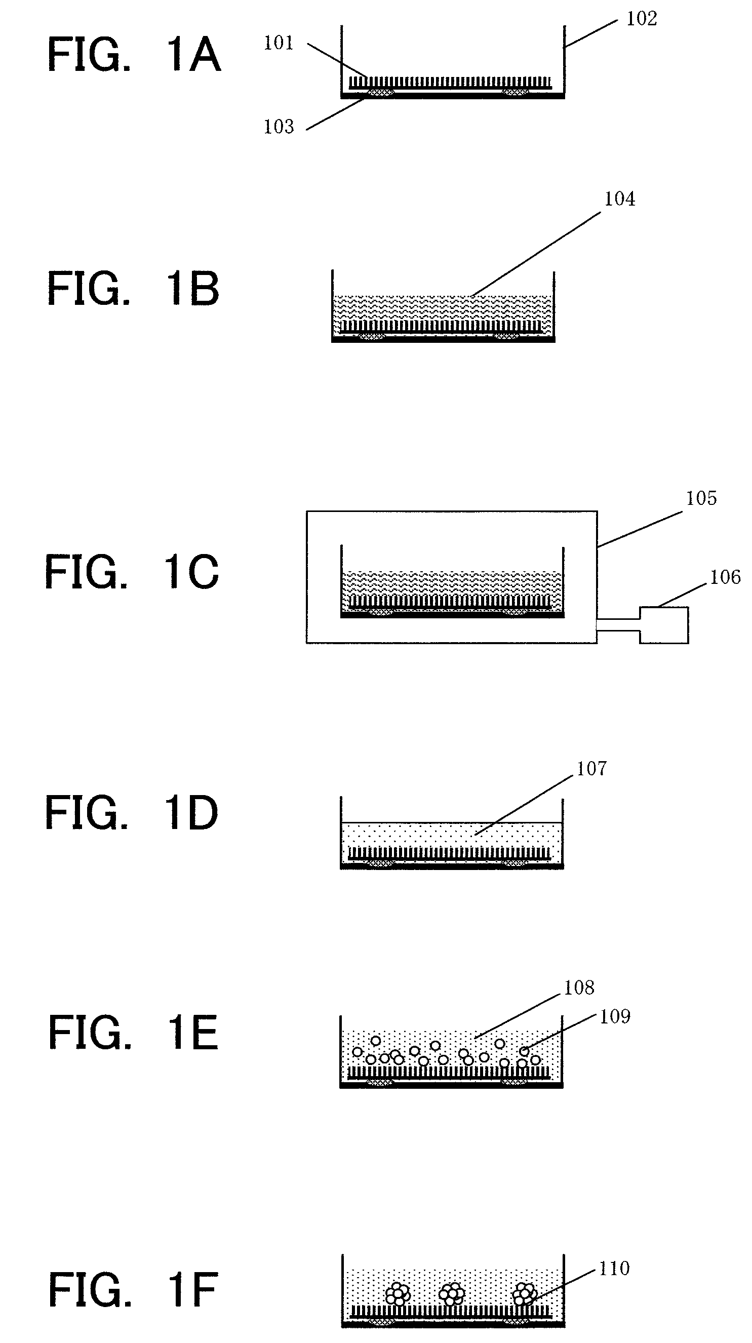
[0028]In Example 1, a step for forming the hepatocyte spheroid on the NP sheet is described.
[0029]
[0030]The preparation of hepatocytes follows an in-situ collagenase perfusion method. The details are as follows. A male Fisher 344 rat (7 to 10 weeks old) is subjected to laparotomy under pentobarbital anesthesia, a catheter is inserted into a portal vein, and a pre-perfusion solution (Hanks' solution without containing Ca2+ and Mg2+ and with containing EGTA) is injected thereinto. At the same time, an inferior vena cava in a lower portion of the liver is incised to release blood. Next, a thoracic cavity is opened, an inferior vena cava entering a right atrium is incised, and the inferior vena cava in the lower portion of the liver is clamped to perform the perfusion. After sufficient blood removal from the liver is confirmed, the perfusion is stopped. A perfusion solution is replaced with a collagenase solution, and the perfusion is performed. In this example, the perfusion is perform...
example 2
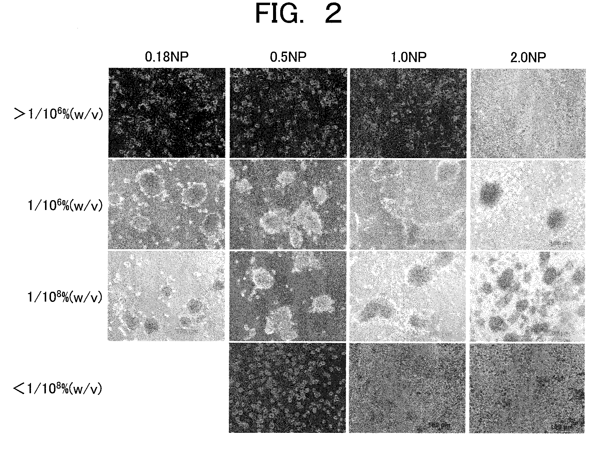
[0039]In Example 2, the effectiveness of the NP hepatocyte spheroid described in the Example 1 is evaluated. Hereinafter, for each evaluation method, an evaluation procedure and an evaluation result are described in this order.
[0040]
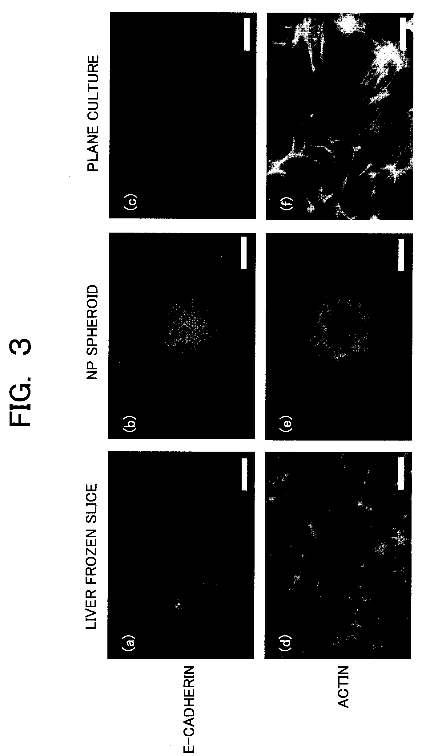
[0041]Immunostaining is performed in order to show that the structure of the formed hepatocyte spheroid is close to the structure of the in-vivo liver tissue. Focused molecules are E-cadherin which is a cell-cell (intercellular) adhesion protein and actin which is a cytoskeletal protein. The E-cadherin is one of membrane proteins playing a role of the cell-cell adhesion binding, and is a protein essential for constructing a tissue having a higher dimensional structure by binding cells with each other. Also, the actin is a cytoskeletal protein providing a physical support to cells when the cells adhere to each other to form the higher dimensional structure. The expression of these two proteins is an index essential to show that the tissue is established a...
example 3
[0060]A configuration of the culture apparatus achieving the automation of the sequence of culture steps described in the Example 1 and its automatic culture steps are described below. FIG. 7 shows an entire configuration diagram of the automatic culture apparatus, FIG. 8 shows details of a decompression unit and a culture unit in the automatic culture apparatus, and FIG. 9 shows a culture automatic flowchart. The conditions are inputted by an input unit, and are displayed on a display unit. The inputted conditions include: an injection amount of the Type I collagen in a Type I collagen injection unit; a pressure inside a decompression chamber in the decompression unit; decompression time; a suction amount of a collagen liquid in a liquid injection / suction unit; an injection amount of a washing liquid; the number of washing times; an injection amount of a cell liquid in a cell suspension injection unit; a culture-unit temperature inside the culture unit; a CO2 concentration inside t...
PUM
| Property | Measurement | Unit |
|---|---|---|
| concentration | aaaaa | aaaaa |
| diameter | aaaaa | aaaaa |
| time | aaaaa | aaaaa |
Abstract
Description
Claims
Application Information
 - R&D
- Intellectual Property
- Life Sciences
- Materials
- Tech Scout
- Unparalleled Data Quality
- Higher Quality Content
- 60% Fewer Hallucinations
Browse by: Latest US Patents, China's latest patents, Technical Efficacy Thesaurus, Application Domain, Technology Topic, Popular Technical Reports.
© 2025 PatSnap. All rights reserved.Legal|Privacy policy|Modern Slavery Act Transparency Statement|Sitemap|About US| Contact US: help@patsnap.com


