
Compositions, devices and methods for SERS and LSPR

a technology applied in the field of sers and lspr, can solve the problem of not immediately diagnosing anthrax, and achieve the effects of large sers enhancement factors, efficient extraction, and stable sers signal

Active Publication Date: 2014-01-14
NORTHWESTERN UNIV +1
View PDF2 Cites 11 Cited by
  • Summary
  • Abstract
  • Description
  • Claims
  • Application Information

AI Technical Summary

Benefits of technology

The present invention is a rapid and portable method and device for detecting pathogenic bacteria (such as anthrax spores) using a low-cost, battery-powered, portable Raman spectrometer. The methods and devices can detect a variety of analytes in different sample types and are especially useful for first-responders. The use of surface-enhanced Raman spectroscopy (SERS) on silver film over nanosphere (AgFON) substrates and atomic layer deposition (ALD) to coat the AgFON surface with a layer of alumina, has improved the binding efficiency and stability of traditional SERS substrates. The speed and sensitivity of the SERS sensor allow for field analysis of potentially harmful environmental samples. Overall, the invention offers a reliable and efficient tool for the detection of pathogens and components of pathogens.

Problems solved by technology

However, the diagnosis of anthrax is not immediate because it takes 1-60 days for anthrax symptoms to appear in humans (Chin, 2000, Control of Communicable Diseases Manual; American Public Health Association: Washington, D.C., pp 20-25).

Method used


Image

  • Compositions, devices and methods for SERS and LSPR
  • Compositions, devices and methods for SERS and LSPR
  • Compositions, devices and methods for SERS and LSPR

Examples


example 1

Microbial Culture

[0091]B. subtilis was purchased from the American Type Culture Collection (Manassas, Va.). Spore cultures were cultivated by spreading the vegetative cells on sterile nutrient agar plates (Fisher Scientific), followed by incubating at 30° C. for 6 days. The cultures were washed from the plates using sterile water and centrifuged at 12 000 g for 10 min. The centrifuging procedure was repeated five times. The lyophilized spores were kept at 2-4° C. prior to use. Approximately 1 g of sample was determined to contain 5.6×10¹⁰ spores by optical microscopic measurements (data not shown). The spore suspension was made by dissolving spores in 0.02 M HNO3 solution and by sonicating for 10 min.

example 2

Extraction of CaDPA from Spores

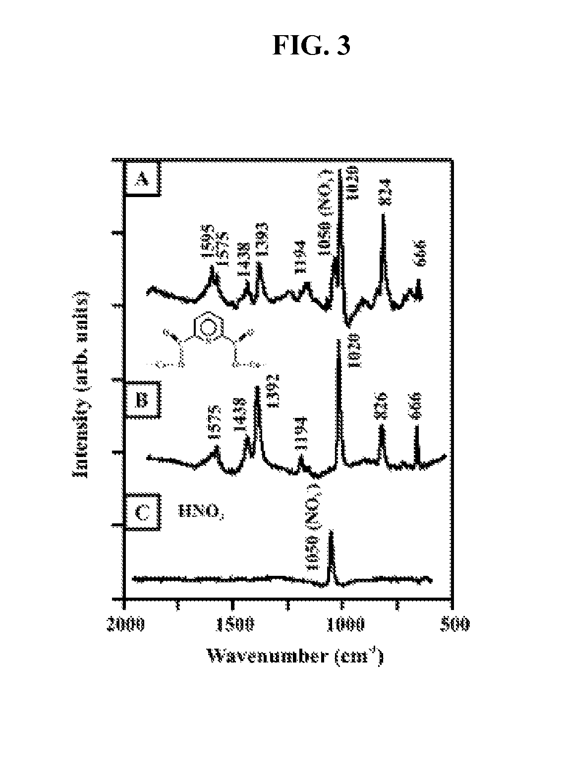
[0092]CaDPA was extracted from spores by sonicating in 0.02 M HNO3 solution for 10 min. This concentration of the HNO3 solution was selected because of its capability of extraction and its benign effect on the AgFON SERS activity. To test the efficiency of this extraction method, a 3.1×10⁻¹³ M spore suspension (3.7×10⁴ spores in 0.2 μL, 0.02 M HNO3) was deposited onto an AgFON substrate (D=600 nm, dm=200 nm). The sample was allowed to evaporate for less than 1 min. A high signal to-noise ratio (S / N) SERS spectrum was obtained in 1 min (FIG. 3A). For comparison, a parallel SERS experiment was conducted using 5.0×10⁻⁴ M CaDPA (FIG. 3B). The SERS spectrum of B. subtilis spores is dominated by bands associated with CaDPA, in agreement with the previous Raman studies on bacillus spores. The SERS spectra in FIG. 3, however, display noticeable differences at 1595 cm-1, which are from the acid form of dipicolinate (Carmona, 1980, Spectrochim. Acta, Part A 36A:...

example 3

AgFON Substrate Fabrication

[0094]Glass substrates were pretreated in two steps; 1) Piranha etch, 3:1 H2SO4 / 30% H2O2 at 80° C. for 1 h, was used to clean the substrate, and 2) base treatment, of 5:1:1 H2O / NH4OH / 30% H2O2 with sonication for 1 h was used to render the surface hydrophilic. Approximately 2 μL of the nanosphere suspension (4% solids) was drop coated onto each substrate and allowed to dry in ambient conditions. The metal films were deposited in a modified Consolidated Vacuum Corporation vapor deposition system with a base pressure of 10−6 Torr (Hulteen and Van Duyne, 1995, J. Vac. Sci. Technol. A 13:1553-1558). The deposition rates for each film (10 Å / s) were measured using a Leybold Inficon XTM / 2 quartz crystal microbalance (QCM) (East Syracuse, N.Y.). AgFON substrates were stored in the dark at room temperature prior to use.

[0095]Alternatively, 2D self-assembled monolayer masks of nanospheres were fabricated by drop-coating approximately 2.54, of undiluted nanosphere sol...


PUM

PropertyMeasurementUnit
diameteraaaaaaaaaa
diameteraaaaaaaaaa
thicknessaaaaaaaaaa

Abstract

The present invention relates to compositions, devices and methods for detecting microorganisms (e.g., anthrax). In particular, the present invention provides portable, surface-enhanced Raman biosensors, and associated substrates, and methods of using the same, for use in rapidly detecting and identifying microorganisms (e.g., anthrax).

Description

[0001]The present invention is a continuation-in-part of, and claims priority to, U.S. Provisional Patent Application Ser. No. 60 / 856,601, filed Nov. 3, 2006 and U.S. patent application Ser. No. 11 / 401,203, filed Apr. 10, 2006, which claims priority to U.S. Provisional Application Ser. No. 60 / 669,727, filed Apr. 8, 2005, each of which is incorporated herein by reference.[0002]This invention was made with government support under DMR-0076097, DMR-0520519, DMR-0520513, EEC-0118025 and CHE-0414554 awarded by the National Science Foundation, under F49620-02-1-0381 awarded by the Air Force Office of Scientific Research MURI, and under DE-FG02-03ER15457 awarded by the U.S. Department of Energy. The government has certain rights in the invention. The United States Government has rights in this invention pursuant to Contract No. W-31-109-ENG-38 between the United States Government and The University of Chicago and / or pursuant to Contract No. DE-AC02-06CH11357 between the United States Gover...

Claims


Application Information

Patent Timeline
Patent Type & Authority Patents(United States)
IPC IPC(8): G01N21/75G01J3/44G01N33/52G01N33/53G01N31/22G01N33/00C12Q1/04
CPCB82Y30/00B82Y15/00G01N21/658C12Q1/04
Inventor VAN DUYNE, RICHARD P.ZHANG, XIAOYUZHAO, JINGWHITNEY, ALYSON V.ELAM, JEFFREY W.SCHATZ, GEORGE C.STAIR, PETER C.ZOU, SHENGLIYOUNG, MATTHEWLYANDRES, OLGA
Owner NORTHWESTERN UNIV
Learn More