Immunoisolation patch system for cellular transplantation

a technology of immunonuclear patch and cellular transplantation, which is applied in the direction of biocide, drug composition, metabolic disorder, etc., can solve the problems of reducing the effect of cellular transplantation, exposing the patient to a wide variety of serious side effects, and causing deleterious effects to the hos

Active Publication Date: 2014-03-18
ENCAPSULIFE
View PDF9 Cites 5 Cited by
  • Summary
  • Abstract
  • Description
  • Claims
  • Application Information

AI Technical Summary

Problems solved by technology

However, a number of technical and logistical challenges have prevented cellular transplantation from working effectively.
This often requires potent immunosuppressive agents having considerable toxicity that can expose the patient to a wide variety of serious side effects.
While immunosuppressive agents increase the chance that the host will accept the cell therapy treatment, it has been well documented that immunosuppressive drugs can cause deleterious effects to the host.
In particular, immunosuppressive agents lower a subject's resistance to infection, make infections harder to treat, and increase the chance of uncontrolled bleeding.
The drugs may also be harmful to the transplanted cells.
However, these designs have limited application, because implantation is highly invasive surgically, device failure is common, and the side effects of device failure are high.
For example, a single breach in macro diffusion devices can cause massive islets death and system failure, and if the breach is sudden, a lethal dose of insulin can be released from the islets, causing death of the host.
Micro-immunoisolation systems, although they overcome many of the problems associated with macro systems, have their own drawbacks.
Moreover, many of these experiments could not be reproduced to acceptable scientific standards.
The lack of experimental control and consistency of those experiments has complicated scientific interpretation and limited their applicability.
Once inside the body cavity, the encapsulated cells could then migrate or diffuse in the body cavity, and may attach to undesirable locations, for example the outer wall of the liver or kidney, which could disrupt the function of those organ, leading to other medical concerns.
Micro-immunoisolation systems also suffer from the effects of gravitational sedimentation, which can lead to undesirable system migration and clumping of encapsulated cells within the body, which can cause a number of undesirable side effects and prevent effective functioning of the implant.
Thus, the promise of immunoprotection of living cells to treat hormone-deficient diseases has not been realized.

Method used


Image

  • Immunoisolation patch system for cellular transplantation
  • Immunoisolation patch system for cellular transplantation
  • Immunoisolation patch system for cellular transplantation

Examples


example 1

Pancreatic Islet Isolation and Evaluation

[0086]For the isolation of pancreatic islets, mongrel canines (20-28 kg body weight) were placed under general anesthesia following an 18-hour fast. A midline laparotomy was performed. The gastroduodenal, splenic and pancreaticoduodenal veins and arteries were isolated and a ligature was placed around each vessel. The main pancreatic duct was identified at the point of duodenal entry and dissected. A ligature was placed around the duct. An 18-gauge angiocath was inserted into the duct and the tip advanced 2-3 mm such that it remained in the main ductal architecture just prior to ductal branching in the pancreas. The catheter was sutured to the duct to secure its position. Immediately prior to harvest, the previously placed vascular ligatures were tightened and the animal was euthanized. The pancreas was transected from all peritoneal and vascular attachments and dissected from the duodenum. Once excised, the pancreas was immediately perfused ...

example 2

Microcapsule Formation

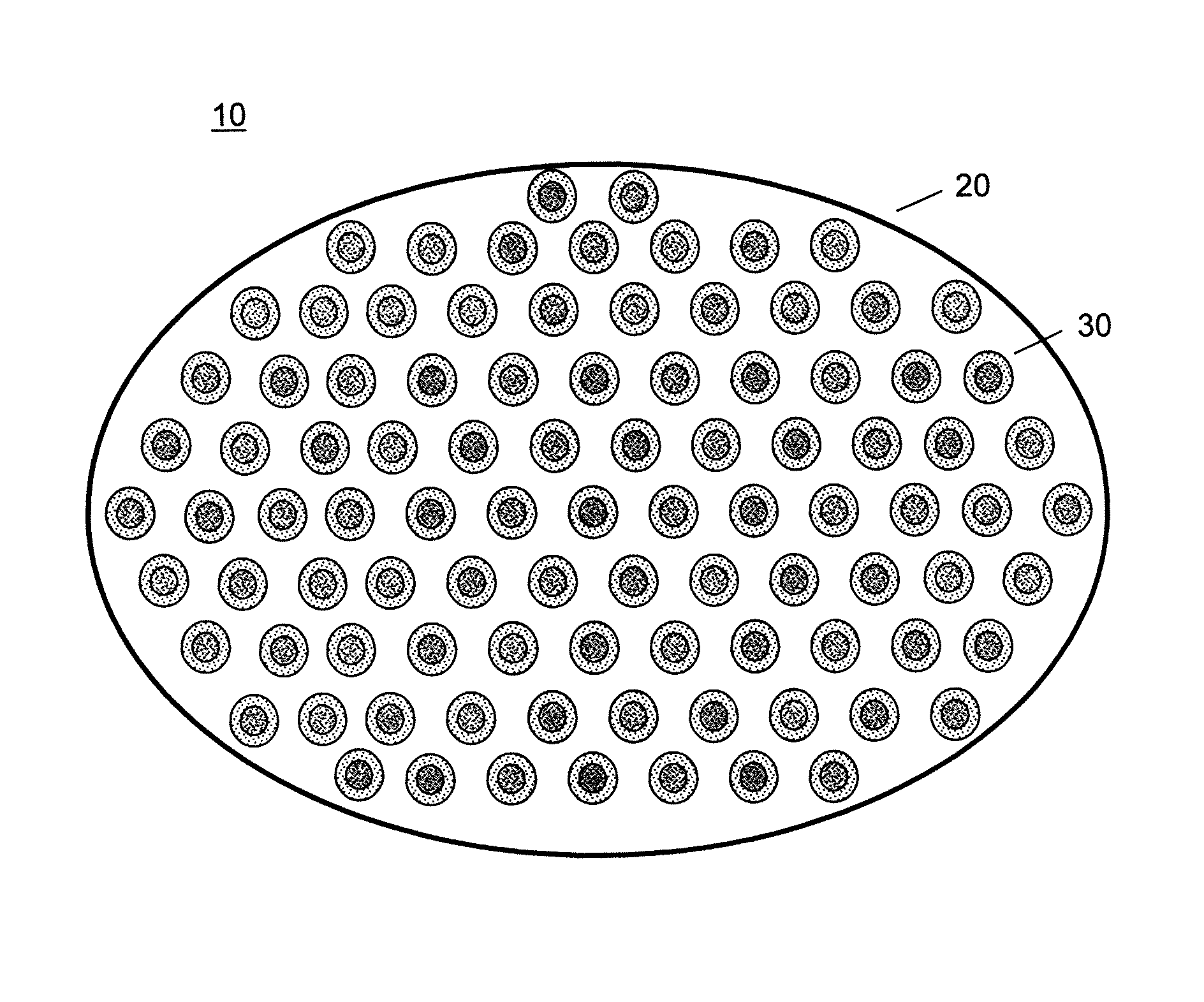
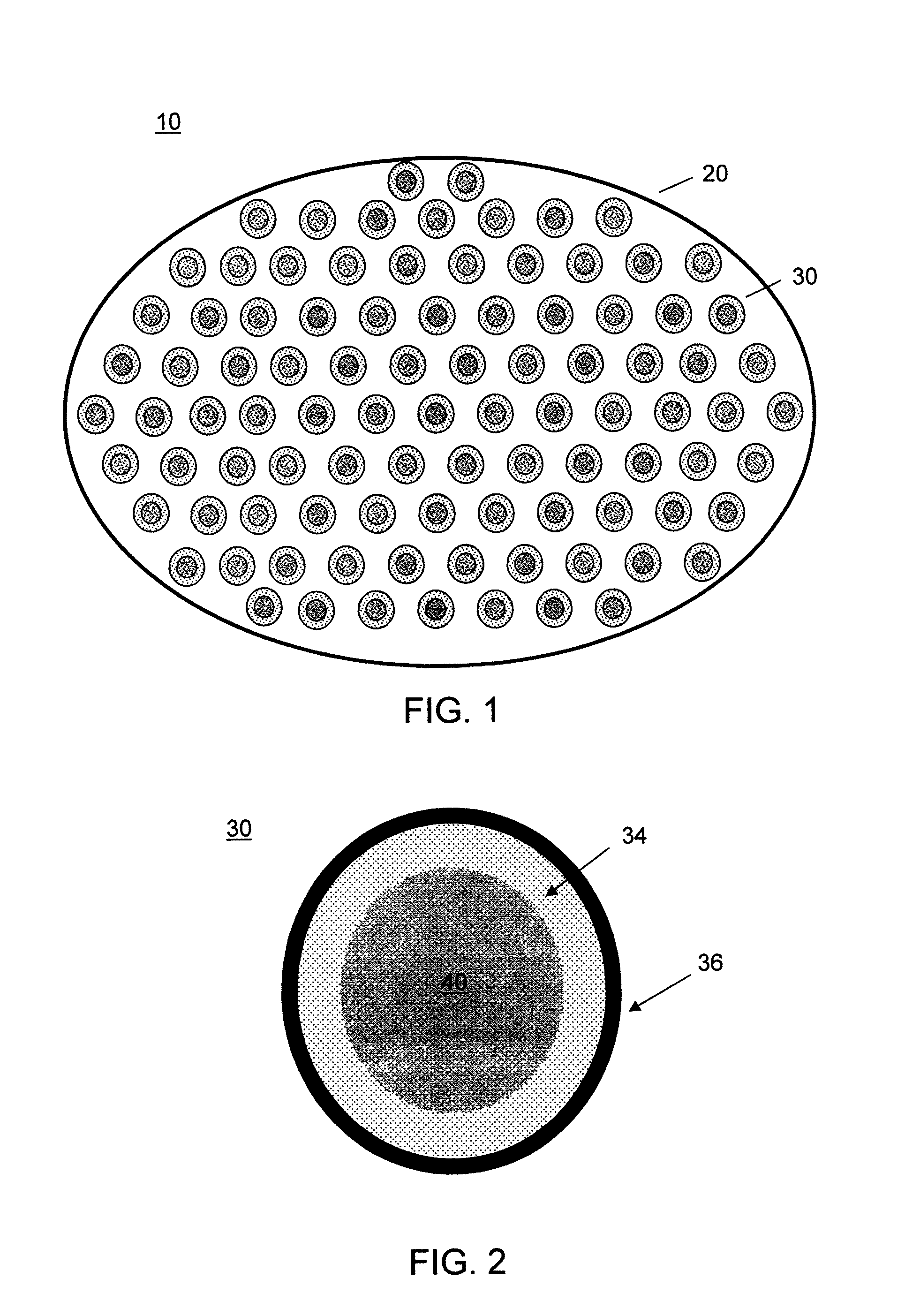
[0091]A series of capsules having a range of permeability (porosity cutoff ranging from 40 kDa-230 kDa, based on dextran exclusion measurement) was developed and characterized. The apparent pore size of the capsular membrane was determined by size exclusion chromatography (SEC) that measures the exclusion of dextran solutes from a column packed with microcapsules. Measuring permeability as a function of polymer component concentration and chemical reaction time allowed adjustment of microcapsule permeability in a controlled manner. Two-membrane microcapsule was formed. The outer membrane with strong ionic bonds formed a thin membrane with narrow pore size distributions. It has a porosity of ˜100 kDa and thickness of ˜1 micron. The inner membrane of this capsule is thicker and its pore size has broad distributions. It has a porosity of approximately 150 kDa and thickness of 20-40 microns.

[0092]Three-membrane microcapsules were also formed, in which the inner mem...

example 3

Encapsulation of Pancreatic Islets and Testing

[0093]Pancreatic islets were isolated as described in Example 1, and following islet isolation, diameter, purity, and viability testing, the islets were cultured for 48-72 hours and encapsulated with a multi-membrane microcapsule of Example 2. The insulin secretory capacity of the free islets and encapsulated islets was then determined in a cell perfusion system.

[0094]Insulin secretion by encapsulated islets was evaluated in a cell perfusion apparatus with a flow rate of 1 ml / minute with RPMI 1640 with 0.1% BSA as a perifusate. Encapsulated islets were perifused with 2 mM glucose for 30 minutes and the column flowthrough discarded. Three minute samples of perifusate were collected during a 30 minute perfusion of 2 mM glucose, a 30 minute perfusion of 20 mM glucose+0.045 mM IBMX (a nutrient), and a 60 minute perfusion of 2 mM glucose. Samples were assayed in duplicate for insulin using Coat-a-Count kits (Diagnostic Products Corporation, L...


PUM


Abstract

An immunoisolation patch system, and particularly a patch system comprising multiple immunoisolation microcapsules, each encapsulating biological material such as cells for transplantation, which can be used in the prophylactic and therapeutic treatment of disease in large animals and humans without the need for immunosuppression.

Description

CROSS REFERENCE TO RELATED APPLICATIONS[0001]The present invention claims priority to U.S. Provisional Patent Application No. 61 / 047,860, filed Apr. 25, 2008, which is incorporated herein by reference in its entirety.STATEMENT REGARDING FEDERALLY-SPONSORED RESEARCH[0002]This invention was made with government support under contract NAG 5-12429 awarded by NASA. The government has certain rights in the invention.FIELD OF THE INVENTION[0003]The present invention relates to an immunoisolation patch system, and particularly a patch system comprising multiple immunoisolation microcapsules, each encapsulating biological material such as cells for transplantation. The present invention also relates to methods of using an immunoisolation patch system for the prophylactic and therapeutic treatment of disease in large animals and humans without the need for immunosuppression.BACKGROUND OF THE INVENTION[0004]Cellular transplantation has recently generated enthusiasm for treating a number of hum...

Claims


Application Information

Patent Timeline
Patent Type & Authority Patents(United States)
IPC IPC(8): A61K35/12A61K9/00A61K9/50
CPCC12N5/0677A61K2035/126C12N2533/32C12N2533/74A61K2035/128C12N2533/30C12N2533/78A61P3/10
Inventor WANG, TAYLOR, G.
Owner ENCAPSULIFE
Why Eureka
Try Eureka