Generation of vascularized human heart tissue and uses thereof

a human heart and vascularization technology, applied in the field of tissue, organ and cell transplantation, can solve the problems of lack of long-term benefits, failure to achieve cardiac cell regeneration, and high cost, and achieve the effect of increasing or decreasing blood flow

Active Publication Date: 2014-07-01
THE GENERAL HOSPITAL CORP
View PDF5 Cites 6 Cited by
  • Summary
  • Abstract
  • Description
  • Claims
  • Application Information

AI Technical Summary

Benefits of technology

[0008]One aspect of the present invention relates to a method to increase the efficiency of differentiation of mbryonic stem (ES) cells, or human induced pluripotent cells (iPSCs) along vasculargenic lineages by contacting the ES cells or iPSCs with VEGF and / or an inhibitor of the TGFβ signaling pathway, such as an ALK5 inhibitor (ALK5i) or the like. Another aspect of the invention relates to increasing the yield of CD31+ cells differentiated from ES cells and / or iPSCs by culturing the cells in a media comprising VEGF or an analogue or functional homologue thereof, and / or an inhibitor of the TGFβ signaling pathway, such as an ALK5 inhibitor (ALK5i) or the like.
[0016]In another embodiment, the human Isl1+ progenitors (which are Isl1+ / Nkx2.5− / KDR−) are differentiated along vasculargenic lineages, e.g., to cardiovascular progenitors, where the cells are differentiated into Isl1+ / CD31+ cells by contacting the cells with at least one of the following; VEGF, VEGF homologue, a inhibitor of TGF-β signalling, an ALK5-inhibitor and the like. In some embodiments, the human Isl1+ progenitors are differentiated along vasculargenic lineages to differentiate into Isl1+ / CD31+ cells by contacting the cells with at least one or more growth factor, selected from the group comprising; the following; vascular endothelial growth factor (VEGF, including, but not limited to VEGF-165), interleukins, fibroblast growth factors, for example, but not limited to, FGF-1 and FGF-2, hepatocyte growth factor, (HGF), endothelins (such as ET-1, ET-2, and ET-3), insulin-like growth factor (IGF-1), angiopoietins (such as Ang-1, Ang-2, Ang-3 / 4), angiopoietin-like proteins (such as ANGPTL1, ANGPTL-2, ANGPTL-3, and ANGPTL-4), platelet-derived growth factor (PDGF), including, but not limited to PDGF-AA, PDGF-BB and PDGF-AB, epidermal growth factor (EGF), endothelial cell growth factor (ECGF), including ECGS, platelet-derived endothelial cell growth factor (PD-ECGF), placenta growth factor (PLGF), and the like. In some embodiments, contacting a population of human Isl1+ progenitors with a VEGF, or a VEGF homologue, or a inhibitor of TGF-β signalling, an ALK5-inhibitor increases the efficiency to differentiate into Isl1+ / CD31+ vasculargenic cells by about at least 1.5-fold, or at least about 2.0-fold, or at least about 3.0-fold, or at least about 4.0-fold, or at least about 5-fold or at least about 6.0-fold or more than 6.0-fold, as compared to in the absence of a VEGF, or a VEGF homologue, or a inhibitor of TGF-β signalling, an ALK5-inhibitor.
[0026]In some embodiments where human ISL1+ progenitors are implanted into an animal subject, the animal can be use as an in vivo humanized model of vascular disease. For example, an animal model which comprises functional vascularized human cardiac tissue, can be used to screen for agents which affect any one, or a combination of viability, functionality, contractibility, differentiation of the human cardiac tissue. As apposed to most in vitro assays or rodent or animal models, the use of the in vivo humanized model of vascular disease has a major advantage over existing assays using cardiac progenitor cells, is that it can be used to monitor or assess the effect of any drug or agent on human cardiac tissue in vivo.
[0036]The use of the in vivo humanized model of vascular disease for identifying agents for effect on human heart function as disclosed herein provides significant advantages over existing method to assess agents on cardiac tissue, because the in vivo humanized model of vascular disease comprises human vascularized cardiac tissue which is formed from ISL1+ primordial cells in vivo, and is properly vascularized and comprises all the desired cell types of heart tissue, including cells of cardiomyocyte phenotypes, endothelial cell phenotypes, smooth muscle phenotypes, as well as characteristics and properties of functional heart tissue. Thus the in vivo humanized model of vascular disease as disclosed herein provides a model of human heart tissue in vivo, which is significantly advantageous over existing cardiac function assays which either are assays using human heart tissue in vitro, or are in vivo models using non-human heart tissue.
[0044]In some instances, reports of ES cell-derived cardiac grafts do not survive longer than three months when in vivo. In some embodiments, the human ISL1+ primordial progenitors are genetically modified to promote survival in response to the stretch stress from contraction. For example, in some embodiments, the human ISL1+ primordial are genetically modified to prolong the survival of humanized vascularized cardiac graft tissue by inhibiting apoptosis, promoting survival pathways, and minimizing immune rejection of the isl1+ progenitors and their differentiated cardiac cells. In some embodiments, the ISL1+ progenitors are genetically modified to express truncated Creb3L2, as disclosed in U.S. Patent Application 61 / 145,208, filed on Jan. 16, 2009, and U.S. patent application Ser. No. 12 / 687,590, filed Jan. 14, 2010 which are both incorporated herein in their entirety by reference.

Problems solved by technology

Nevertheless, the demand for transplantation exceeds the availability of donor hearts.
However, their low frequency of cardiac differentiation (Murry et al., 2004) and lack of long-term benefits fail to achieve cardiac cell regeneration (Fazel et al., 2006, 2008).
Disruption of development, differentiation or maturation of any of these components can lead to arrhythmias such as sinus arrest, AV block, ventricular tachycardia and sudden death (Bruneau et al., 2001).
Unraveling the mechanisms for human heart cell lineage diversification has been hampered by the lack of genetic tools to purify early cardiac progenitors and define their developmental potential1-4.

Method used


Image

  • Generation of vascularized human heart tissue and uses thereof
  • Generation of vascularized human heart tissue and uses thereof
  • Generation of vascularized human heart tissue and uses thereof

Examples


example 1

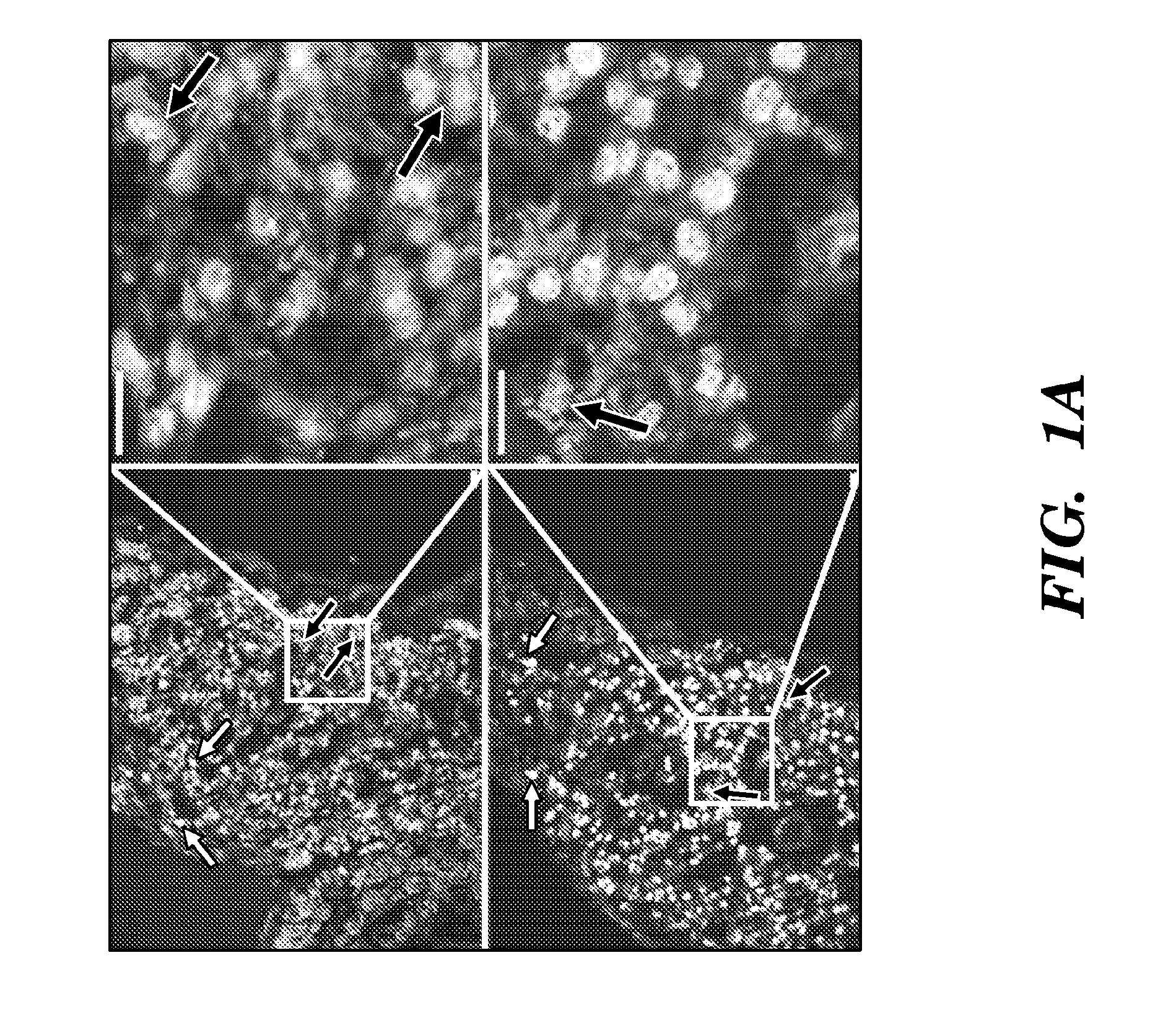
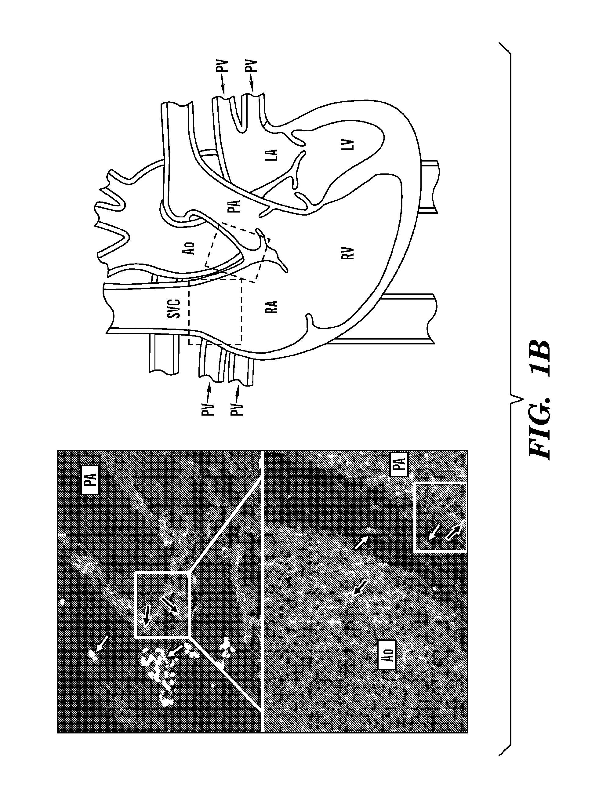
[0484]To localize ISL1+ heart cells during human cardiogenesis, the inventors employed immunofluorescence microscopy in serial sections of human fetal hearts at 11 and 18 weeks of gestation. ISL1+ cells were found in the right atrium (RA) and outflow tract (OFT), both second heard field (SHF)-derived structures (FIG. 1), as well as in the left atrial wall and appendage. In particular, the inventors demonstrated that in vivo expression of ISL1 in the human fetal heart using hematoxylin eosin staining of left atrium and left atrium appendage of a human fetal heart at 11 weeks of gestation (data not shown). The inventors also performed double immunostaining of sections of the human heart from RA region with antibodies directed to ISL1 and antibodies directed to one or more of cTNT, SMMHC, PECAM1, NKX2.5 and detected double immunostained cells positive for ISL1 and PECAM1 (data not shown).

[0485]Thus, the inventors demonstrated that ISL1+ cells in the human fetal heart are found in patte...

example 2

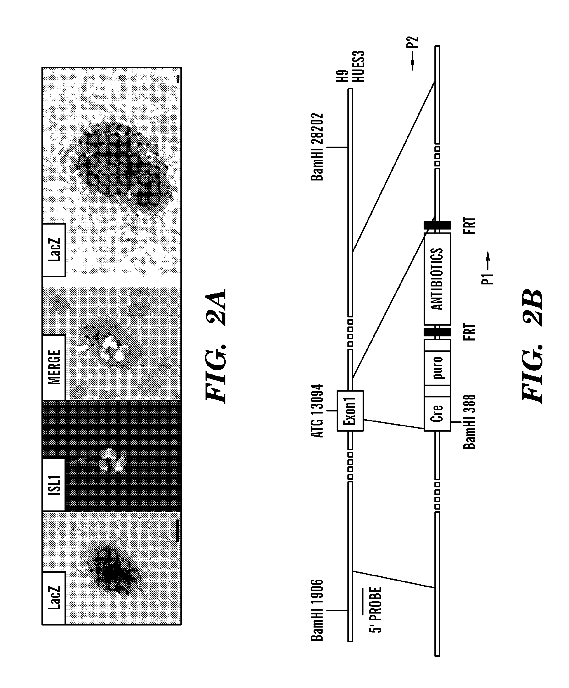
[0489]To obtain purified populations of ISL1+ cardiac progenitors, the inventors isolated DsRed+ cells by FACS on EB day 8 (FIG. 3B). RT-PCR analysis revealed that the expression of cardiac markers ISL1 and NKX2-5 is 30- and 8-fold higher in DsRed+ cells than that in DsRed− population, respectively (FIG. 3C). Interestingly, both FACS and RT-PCR analyses demonstrated that DsRed+ cells did not express KDR when isolated on EB day 8 (FIG. 7A-7B).

[0490]To assess the differentiation potential of human ISL1+ progenitors, the inventors performed clonal assays on single DsRed+ cells. The inventors them isolated DsRed+ cells from day 8 EBs and plated them at low density on mouse embryonic fibroblast (MEF) feeders. Colonies of DsRed+ cells were observed within seven days at a cloning efficiency of 0.2-0.5%. The inventors demonstrated that FACS isolated DsRed+ cells from day 8 human ISL1-cre DsRed EBs formed colonies on MEF feeders after 7-day and 14-days (data not shown). The inventors perform...

example 3

[0500]Cardiovascular disease is the leading cause of death in the U.S., and will be the primary cause of mortality in developing countries by 2010, as estimated by the WHO. Nevertheless, the demand for transplantation exceeds the availability of donor hearts. In this regard, cardiac regeneration has recently become an active area of research. Over the past few years, numerous reports demonstrate cardiac progenitors from diverse fetal and adult tissues outside the cardiovascular system, including adipose tissues, amniotic fluid, bone marrow, placenta, skeletal muscle, and testes (Franco et al., 2007). However, their low frequency of cardiac differentiation (Murry et al., 2004) and lack of long-term benefits fail to achieve cardiac cell regeneration (Fazel et al., 2006, 2008). Bone marrow cells, for instance, may improve the function of the infarcted heart mainly by promoting angiogenesis or cell survival without cardiac muscle regeneration (Fazel et al., 2006, 2007). A recently disco...


PUM

PropertyMeasurementUnit
concentrationaaaaaaaaaa

Abstract

The present invention relates to the generation of vascularized human heart tissue from human primordial Islet1-positive (ISL1+) progenitors, and more particularly the generation of vascularized human heart tissue from human primordial Islet1+ cardiovascular stem cells which are positive for markers ISL1+ / NKX2.5− / KDR−. One aspect of the invention relates to isolation of human ISL1+ primordial cells from human pluripotent cells, such as human ES cells or other human pluripotent stem cell sources, wherein the human ISL1+ primordial cells can differentiate into three different lineages; cardiomyocyte lineages, endothelial lineages and smooth muscle lineages. Another aspect relates to use and implantation of the human primordial ISL1+ progenitors into an animal model to generate human vascularized heart tissue, and more particularly, the production of an in vivo humanized model of vascular disease. One embodiment relates to the use of an in vivo humanized model of vascular disease as an assay, for example to assess drug toxicity and / or identify agents which increase and decrease coronary blood flow to the human vascularized heart tissue. Another embodiment relates to the therapeutic use of human primordial ISL1+ progenitors, for example, in one embodiment the invention provides methods for the treatment cardiovascular disorders and / or congenital heart disease in a subject comprising transplanting into subjects vascularized human heart tissue generated from human ISL1+ progenitors.

Description

CROSS REFERENCE TO RELATED APPLICATION[0001]This Application is a 35 U.S.C. §371 National Phase Entry Application of International Application No. PCT / US2010 / 038134 filed Jun. 10, 2010, which designates the U.S., and which claims benefit under 35 U.S.C. §119(e) to U.S. Provisional Application 61 / 185,752, filed, Jun. 10, 2009 and U.S. Provisional Application 61 / 256,960, filed, Oct. 31, 2009, the entire contents of each are incorporated herein by reference.FIELD OF THE INVENTION[0002]The present invention relates to the field of tissue, organ and cell transplantation. Methods and compositions of production of human vascularized cardiac tissue are provided as well as an in vivo model of human cardiovascular disease and an in vivo assay for monitoring cardiac development, as well as identifying agents affecting human cardiovascular function.BACKGROUND OF THE INVENTION[0003]Cardiovascular disease is the leading cause of death in the U.S., and will be the primary cause of mortality in dev...

Claims


Application Information

Patent Timeline
Patent Type & Authority Patents(United States)
IPC IPC(8): A01N63/00G01N33/53A61K35/12
CPCC12N5/0662A61K35/12C12N5/0657C12N2501/15C12N2501/165C12N2501/415C12N2506/02A61P9/00
Inventor CHIEN, KENNETH R.BU, LEIJIANG, XINLUI, KATHY OI LANCHMIELOWIEC, JOLANTA
Owner THE GENERAL HOSPITAL CORP
Why Eureka
Try Eureka