Transapical mitral valve repair device

a technology repair device, which is applied in the field of transapical mitral valve repair device, can solve the problems of affecting the proper functioning of one or more of the valves of the heart, prolapse and regurgitation, and obstruction of blood flow, and achieves the effect of facilitating mitral valve repair

Active Publication Date: 2014-10-07
UNIV OF MARYLAND
View PDF96 Cites 95 Cited by
  • Summary
  • Abstract
  • Description
  • Claims
  • Application Information

AI Technical Summary

Benefits of technology

[0023]It is another object of the disclosure to enable chordal replacement with ePTFE. A related object of the present disclosure is to provide a chordal replacement that facilitates mitral valve repair.
[0024]Another object of the disclosure is to provide a method and device for transapical mitral valve repair that uses a small incision. A related object of the disclosure is to provide a method that does not require a Sternotomy. Another related object of the disclosure is to provide a method that does not require cardiopulmonary bypass or aortic manipulation.
[0027]These and other objects of the present disclosure are accomplished by providing a device for minimally invasive repair of a defective heart valve while the heart is beating. The heart can be accessed through the apex or a point lateral / near to the apex with a small-diameter shafted instrument. The instrument might be a needle or a catheter. Using ultrasound guidance (real-time transesophagael echocardiography), the shafted instrument is inserted through an access port at the apex (or near the apex) and the instrument is guided to make contact with the mitral valve leaflet at the location where the operator has decided that a neochord should be inserted. Typically, this would be the body of the anterior or posterior leaflet in a location where the valve has prolapsed as a result of a broken or elongated chord. The instrument punctures the apex of the heart and travels through the ventricle. The tip of the instrument rests on the defective valve and punctures the valve leaflet. The instrument then inserts either a suture or a suture / guide wire combination, securing the top of the leaflet to the apex of the heart with an artificial chordae. A resilient element or shock absorber mechanism adjacent to the outside of the apex of the heart minimizes the linear travel of the instrument in response to the beating of the heart or opening / closing of the valve.

Problems solved by technology

Various disease processes can impair the proper functioning of one or more of the valves of the heart.
Additionally, damage to the ventricle from prior heart attacks (i.e., myocardial infarction secondary to coronary artery disease) or other heart diseases (e.g., cardiomyopathy) can distort the valve's geometry causing it to dysfunction.
However, the vast majority of patients undergoing valve surgery, such as mitral valve surgery, suffer from a degenerative disease that causes a malfunction in a leaflet of the valve, which results in prolapse and regurgitation.
One possible malfunction, valve stenosis, occurs when a valve does not open completely and thereby causes an obstruction of blood flow.
Typically, stenosis results from buildup of calcified material on the leaflets of the valves causing them to thicken and thereby impairing their ability to fully open and permit adequate forward blood flow.
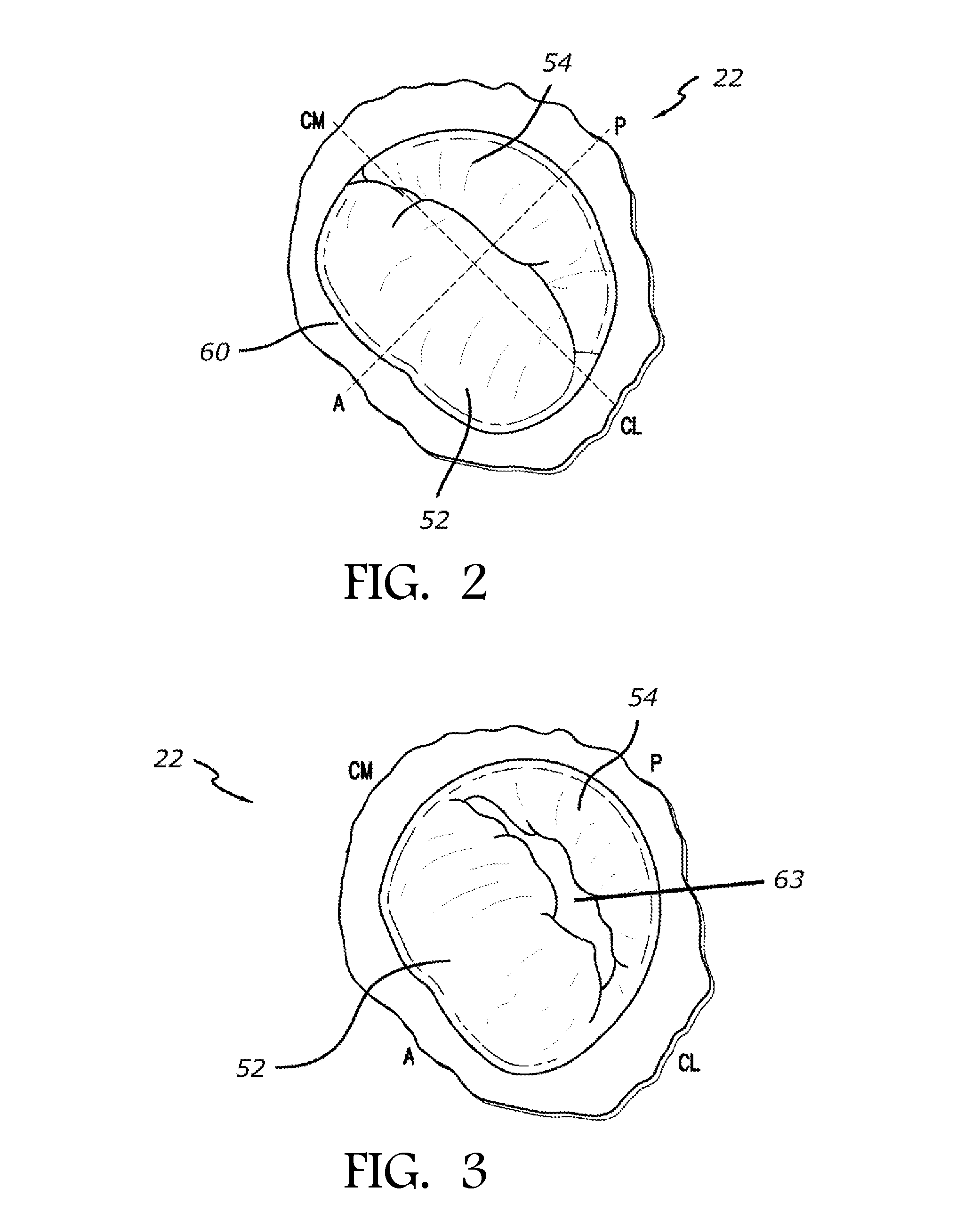
Another possible malfunction, valve regurgitation, occurs when the leaflets of the valve do not close completely thereby causing blood to leak back into the prior chamber.
A Carpentier type I malfunction involves the dilation of the annulus such that normally functioning leaflets are distracted from each other and fail to form a tight seal (i.e., do not coapt properly).
This is the most common cause of mitral regurgitation, and is often caused by the stretching or rupturing of chordae tendineae normally connected to the leaflet.
A Carpentier's type III malfunction involves restriction of the motion of one or more leaflets such that the leaflets are abnormally constrained below the level of the plane of the annulus.
As set forth above, there are several different ways a leaflet may malfunction, which can thereby lead to regurgitation.
Both valve stenosis and valve regurgitation increase the workload on the heart 10 and may lead to very serious conditions if left un-treated; such as endocarditis, congestive heart failure, permanent heart damage, cardiac arrest, and ultimately death.
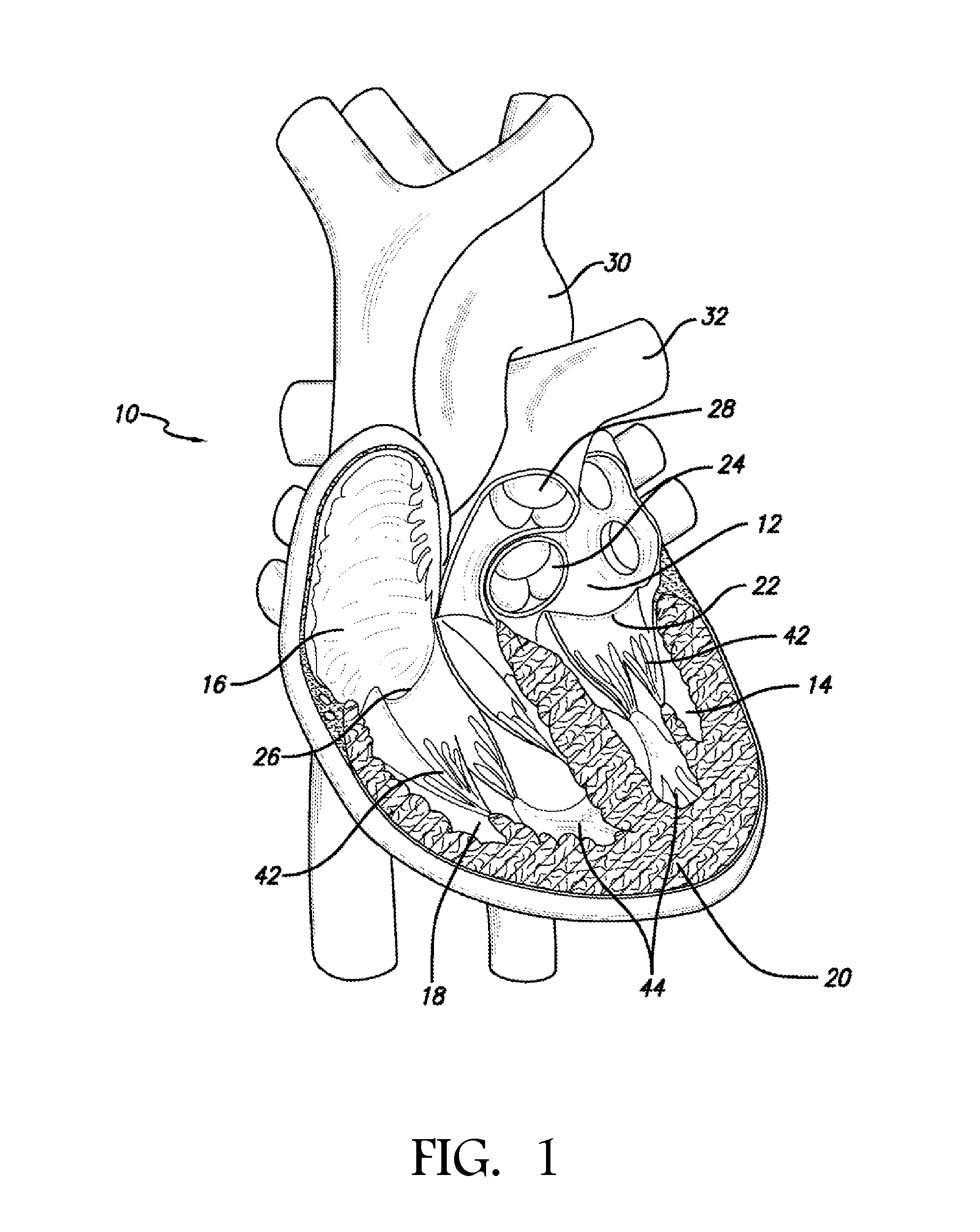
Since the left heart is primarily responsible for circulating the flow of blood throughout the body, malfunction of the mitral valve 22 or tricuspid valve 26 is particularly problematic and often life threatening.
Accordingly, because of the substantially higher pressures on the left side of the heart, left-sided valve dysfunction is much more problematic.
Deformation of the leaflets, as described above, prevents the valves from closing properly and allows for regurgitation or back flow from the ventricle into the atrium, which results in valvular insufficiency.
Bioprosthetic valves have limited durability.
Moreover, prosthetic valves rarely function as well as the patient's own valves.
Additionally, there is an increased rate of survival and a decreased mortality rate and incidence of endocarditis for repair procedures.
Further, because of the risk of thromboembolism, mechanical valves often require further maintenance, such as the lifelong treatment with blood thinners and anticoagulants.
However, because of the complex and technical demands of the repair procedures, the overall repair rate in the United States is only around 50%.
Conventional techniques for repairing a cardiac valve are labor-intensive, technically challenging, and require a great deal of hand-to-eye coordination.
They are, therefore, very challenging to perform, and require a great deal of experience and extremely good judgment.
Additionally, leaflet sparing procedures for correcting regurgitation are just as labor-intensive and technically challenging, if not requiring an even greater level of hand-to-eye coordination.
These procedures, however, adversely affect almost all of the organ systems of the body and may lead to complications, such as strokes, myocardial “stunning” or damage, respiratory failure, kidney failure, bleeding, generalized inflammation, and death.
The risk of these complications is directly related to the amount of time the heart is stopped (“cross-clamp time”) and the amount of time the subject is on the heart-lung machine (“pump time”).
Furthermore, the conventional methods currently being practiced for the implantation of the artificial chordae are particularly problematic.
Because the conventional approach requires the heart to be stopped (e.g., via atriotomy) it is difficult to accurately determine, assess, and secure the appropriate chordal length.
Using conventional techniques, it is very difficult to ensure that the chordae are of the correct length and are appropriately spaced inside the ventricle to produce a competent valve.

Method used


Image

  • Transapical mitral valve repair device
  • Transapical mitral valve repair device
  • Transapical mitral valve repair device

Examples


Embodiment Construction

[0054]In accordance with the methods of embodiments herein, the heart may be accessed through one or more openings made by a small incision(s) in a portion of the body proximal to the thoracic cavity, for instance, in between one or more of the ribs of the rib cage, proximate to the xyphoid appendage, or via the abdomen and diaphragm. Access to the thoracic cavity may be sought so as to allow the insertion and use of one or more thorascopic instruments, while access to the abdomen may be sought so as to allow the insertion and use of one or more laparoscopic instruments. Insertion of one or more visualizing instruments may then be followed by transdiaphragmatic access to the heart. Additionally, access to the heart may be gained by direct puncture (i.e., via an appropriately sized needle, for instance an 18 gauge needle) of the heart from the xyphoid region. Access may also be achieved using percutaneous means. Accordingly, the one or more incisions should be made in such a manner a...


PUM


Abstract

Methods and devices for repairing a cardiac valve. A minimally invasive procedure includes creating an access in the apex region of the heart through which one or more instruments may be inserted. The device can implant artificial heart valve chordae tendineae into cardiac valve leaflet tissues to restore proper leaflet function and prevent reperfusion. The device punctures the apex of the heart and travels through the ventricle. The tip of the device rests on the defective valve and punctures the valve leaflet. A suture or a suture / guide wire combination is inserted, securing the top of the leaflet to the apex of the heart. A resilient element or shock absorber mechanism adjacent to the outside of the apex of the heart minimizes the linear travel of the device in response to the beating of the heart or opening / closing of the valve.

Description

RELATED APPLICATIONS[0001]This application is a continuation of and claims priority to International Application No: PCT / US2012 / 043761, filed Jun. 22, 2012, entitled “Transapical Mitral Valve Repair Device,” which claims priority to and the benefit of U.S. Provisional Application No. 61 / 501,404, filed Jun. 27, 2011, entitled “Transapical Mitral Repair with Pre-Formed Knot,” and U.S. Provisional Application No. 61 / 550,772, filed Oct. 24, 2011, entitled “Transapical Mitral Valve Repair Device,” the disclosures of which are incorporated herein by reference in their entirety.BACKGROUND[0002]1. Field of the Disclosure[0003]The disclosure herein relates to methods and devices for performing cardiac valve repairs, and more particularly, the disclosure relates to methods and devices for performing minimally invasive mitral or tricuspid valve repairs using of PTFE neochords through a minimally invasive incision, while the heart is beating.[0004]2. Description of the Background[0005]As illust...

Claims


Application Information

Patent Timeline
Patent Type & Authority Patents(United States)
IPC IPC(8): A61B17/04A61F2/08A61B17/00A61F2/24
CPCA61B17/0401A61B17/0485A61F2/2457A61B2017/0404A61B17/0469A61B2017/00783A61B2017/0409A61B2017/00243A61F2/2466A61B2017/0464A61B2017/0406A61B2017/0417A61B2017/047A61B2017/06171A61B17/062A61B17/06166A61B17/06066
Inventor GAMMIE, JAMESPATEL, RAHULGHOREISHI, MEHRDAD
Owner UNIV OF MARYLAND
Why Eureka
Try Eureka