

Detection of an amplification reaction product using pH-sensitive dyes
a technology of amplification reaction product and ph-sensitive dye, which is applied in the direction of fluorescence/phosphorescence, instruments, biochemistry apparatus and processes, etc., can solve the problems of long incubation time, inability to detect specific instruments, and inability to detect subtle changes,
- Summary
- Abstract
- Description
- Claims
- Application Information
AI Technical Summary
Benefits of technology
Problems solved by technology
Method used
Image

Examples
example 1
Detection of LAMP Amplification Using a pH-Sensitive Visual Dye
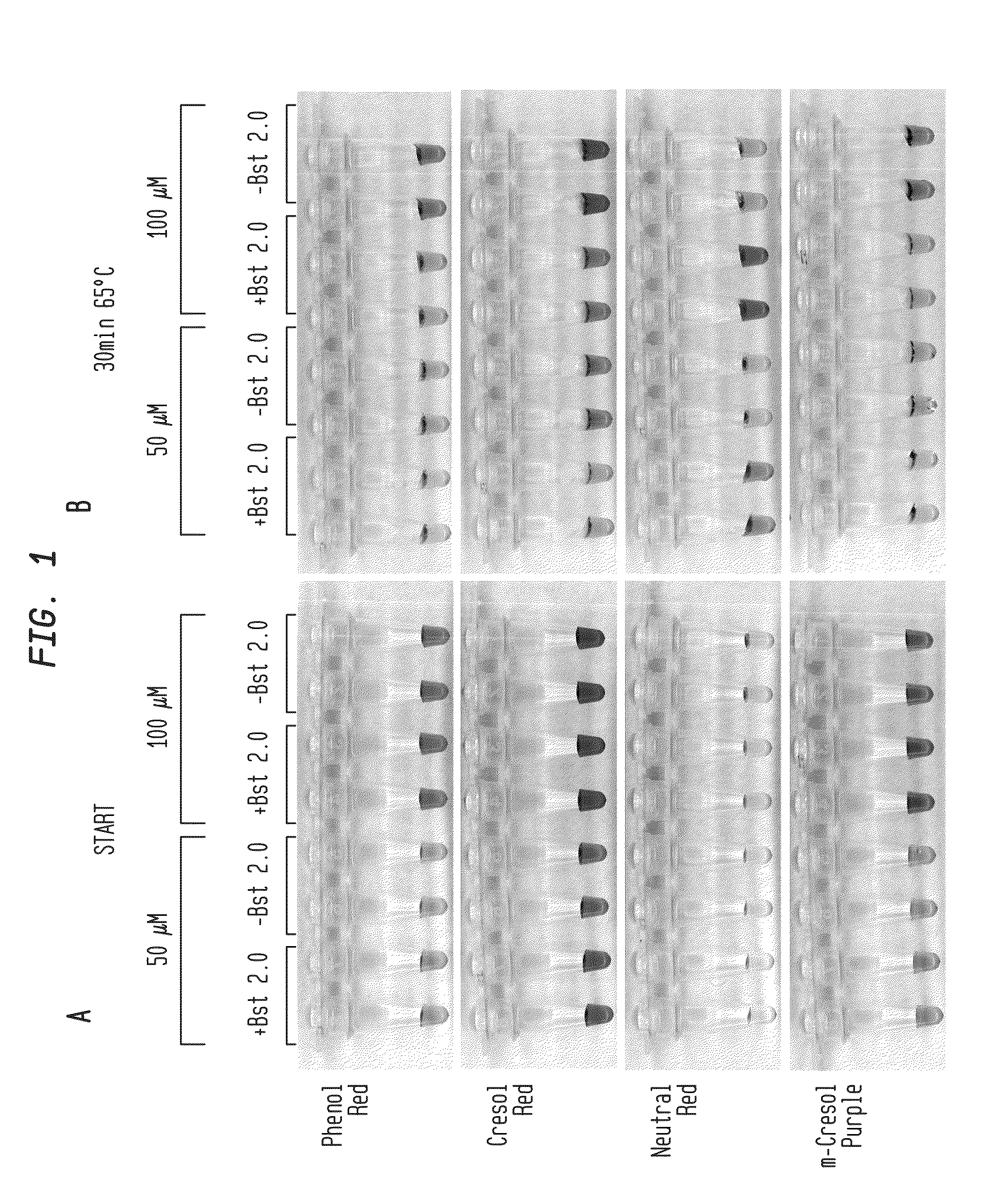
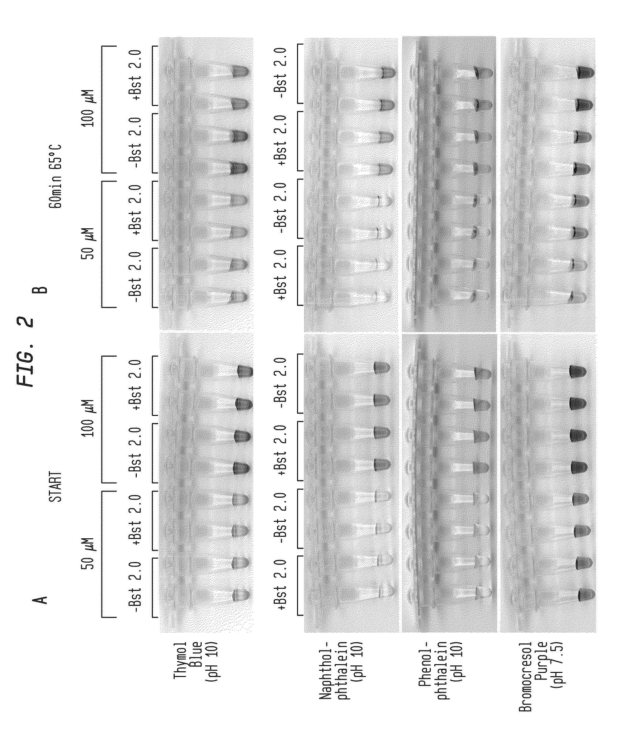
[0069]LAMP reactions were performed with a buffer-free reaction solution: 10 mM (NH4)2SO4, 50 mM KCl, 8 mM MgSO4, 1.4 mM dNTPs, 0.1% Tween-20, pH 7.5-10. Final buffer concentration was 0.026 mM-0.4 mM Tris from enzyme storage buffer carryover.
[0070]Reactions were performed with primers for lambda phage DNA amplicon and 5 ng of lambda DNA (FIG. 1A-B, FIG. 2A-B; New England Biolabs, Ipswich, Mass.). Reactions were incubated for 30-60 minutes at 65° C. with either 50 μM or 100 μM pH indicator as shown in the presence or absence of DNA polymerase (Bst 2.0). Color change occurred only in the presence of DNA polymerase, indicating that amplification produced sufficient pH drop for visual identification.
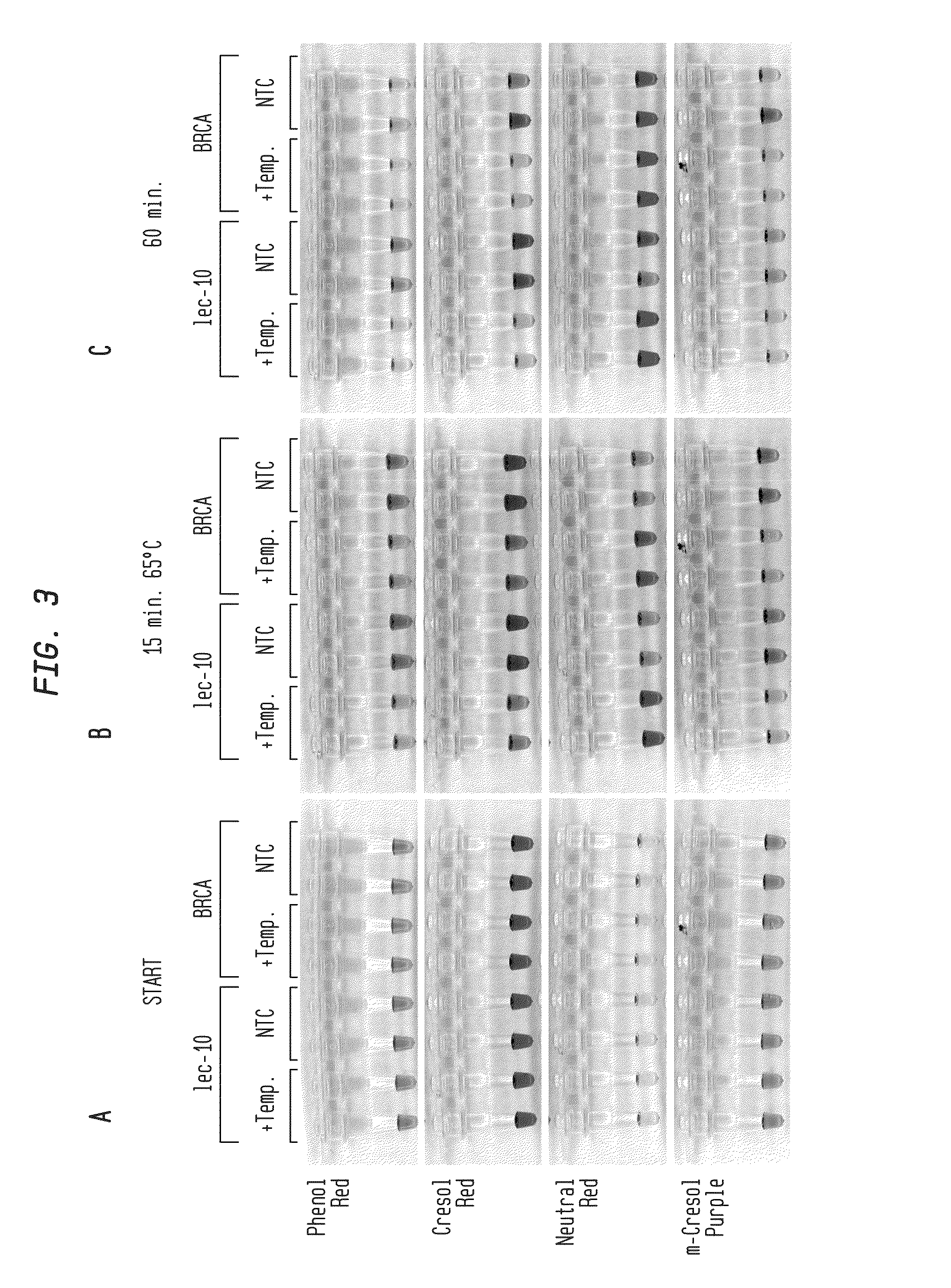
[0071]In FIG. 3A-C, LAMP reactions were performed in buffer-free reaction solution with primers for either C. elegans lec-10 or human BRCA1 sequence targets. Reactions contained 82.5 ng C. elegans DNA, 100 ng HeLa DNA (+Temp) or...
example 2
Detection of PCR Amplification Using a pH-Sensitive Visual Dye
[0074]The PCR reaction was performed in 50 mM KCl and 2.25 mM MgCl2 using 500 nM each of the forward and reverse primers that amplify a 1.287 kb fragment from pAII17 plasmid DNA, 400 μM each of four dNTPs, 100 μM phenol red, 0.025 μl of 1M KOH, 1.875 U of Taq DNA polymerase in 25 μl. The PCR reaction was performed at 95° C. for 2 minutes, 36 cycles of 95° C. for 10 seconds, 62° C. for 15 seconds, 68° C. for 30 seconds. Before PCR cycling, all tubes, either with or without DNA template, had the same pink color. At the end of the PCR reaction, the triplicate reactions (labeled 1, 2 and 3; FIG. 5) that had DNA template changed color from pink to yellow while the reactions without DNA template (labeled 4, 5, and 6; FIG. 5) remained pink. DNA synthesis in the reactions containing template was confirmed using real-time PCR machine and agarose gel electrophoresis. Thus, the color change provided a reliable visual indicator for s...
example 3
Visual Detection of Plasmid DNA in Bacterial Colonies
[0076]PCR reactions were performed in the presence of phenol red to identify E. coli colonies that were transformed to carry a specific plasmid DNA. A small portion of each colony was suspended in 10 μl water and 1 μl was added in the PCR reaction, which was performed as described in Example 2. Six colonies were tested with three colonies (1-3) from a plate that carries the same plasmid as used in the positive control (+) and three colonies (a-c) from a bacterial plate containing an unrelated plasmid. As in the positive control, the tubes that contained the target plasmid DNA changed color from pink to yellow (FIG. 6). The tubes that contained the unrelated plasmid remained pink just like the tube without any template (−). Thus, the color change in these PCR reactions allowed determination of colonies containing a specific plasmid DNA. This approach avoided a conventional step of using agarose gel electrophoresis to determine the ...
PUM
| Property | Measurement | Unit |
|---|---|---|
| pH | aaaaa | aaaaa |
| pH | aaaaa | aaaaa |
| pH | aaaaa | aaaaa |
Abstract
Description
Claims
Application Information