
Systems, devices and methods for providing therapy to an anatomical structure using high frequency pressure waves and/or cryogenic temperatures

a technology of high frequency pressure wave and anatomical structure, applied in the field of systems, devices and methods for providing therapy to anatomical structure using high frequency pressure wave and/or cryogenic temperature, can solve the problems of large patient population that cannot tolerate the prescribed medications or are not considered candidates, and cannot offer mechanical therapy to these patients, so as to reduce the number of different devices needed and reduce the cost of the procedure

Active Publication Date: 2016-10-25
ARTHROCARE
View PDF60 Cites 3 Cited by
  • Summary
  • Abstract
  • Description
  • Claims
  • Application Information

AI Technical Summary

Benefits of technology

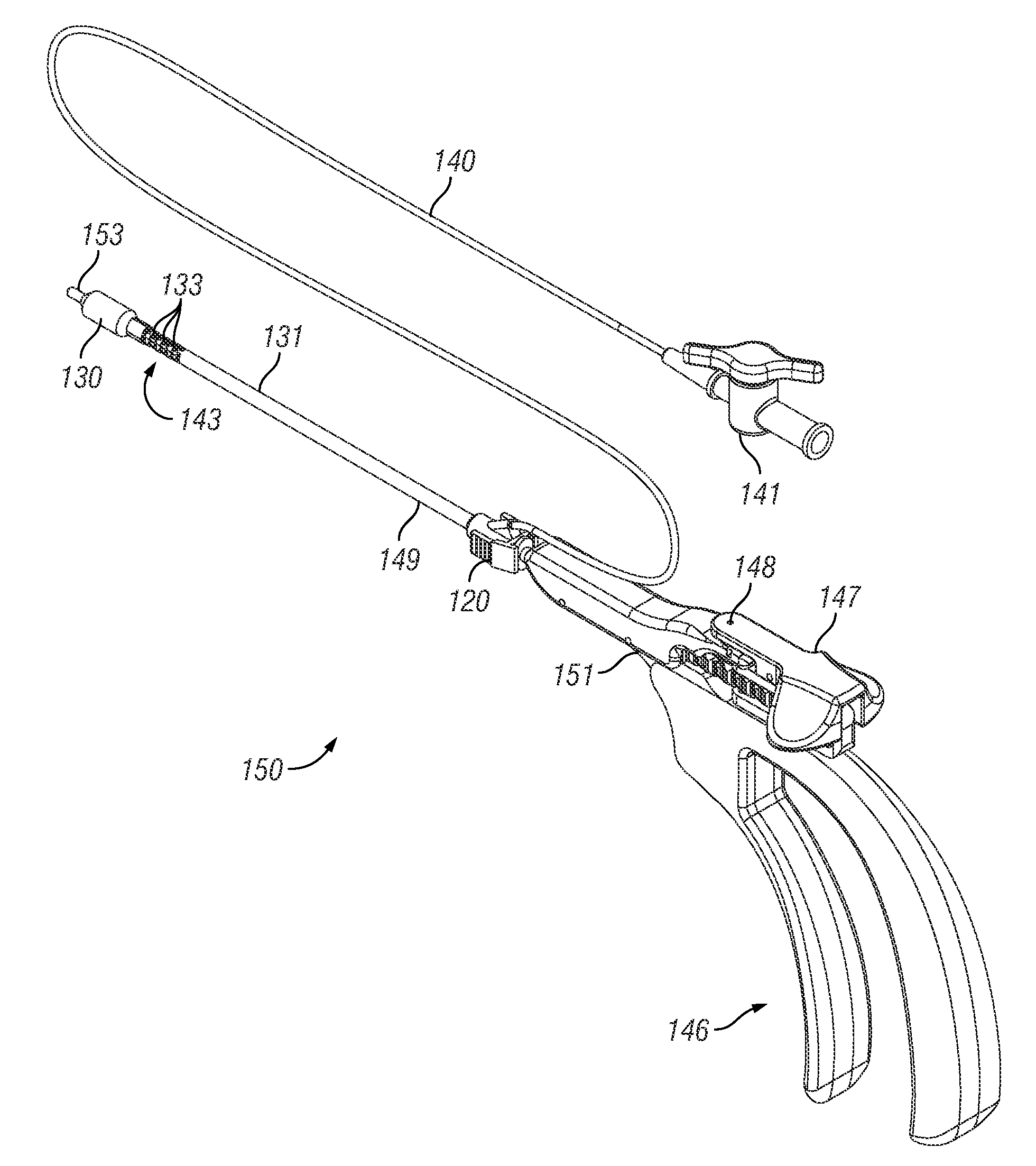
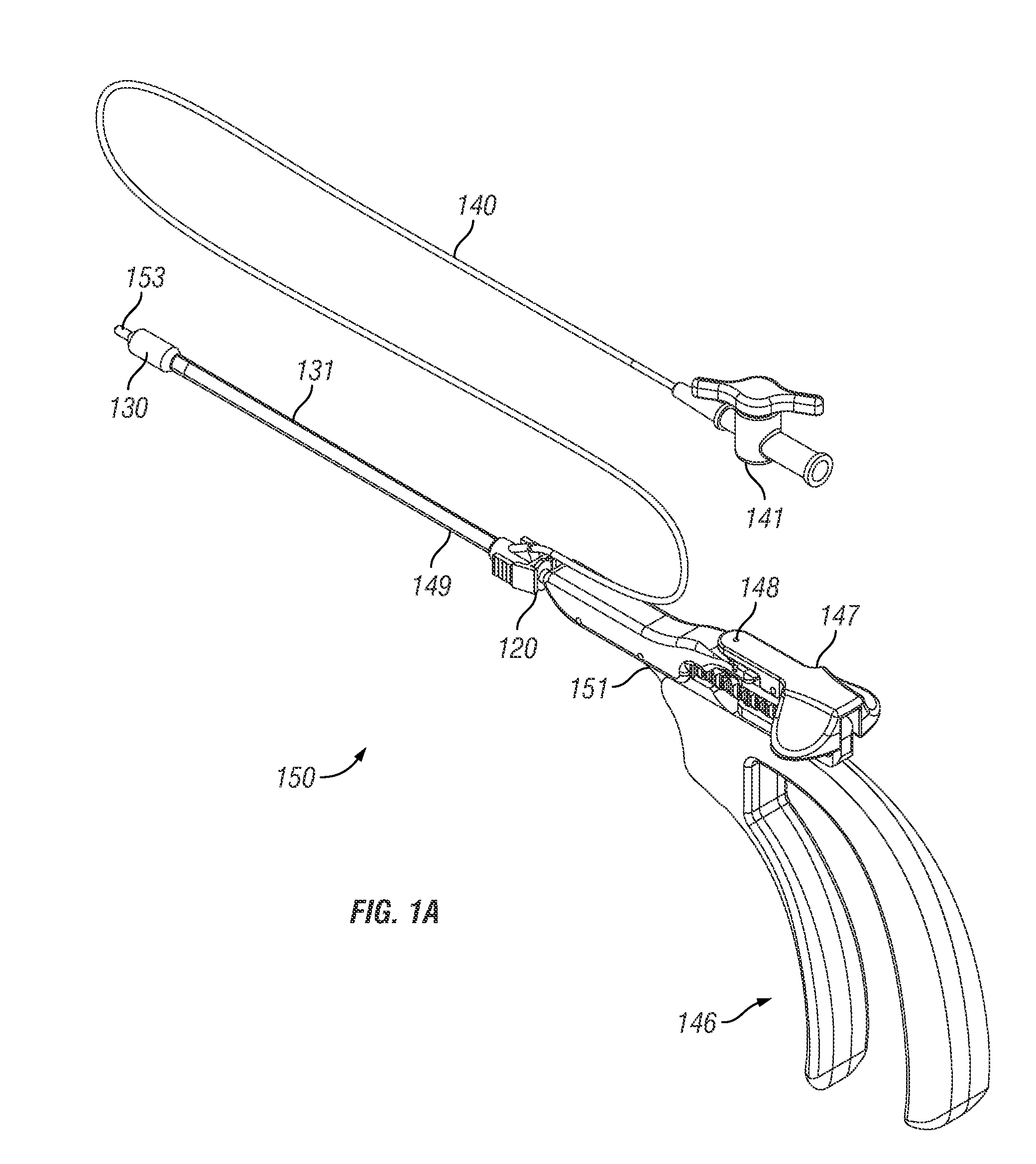
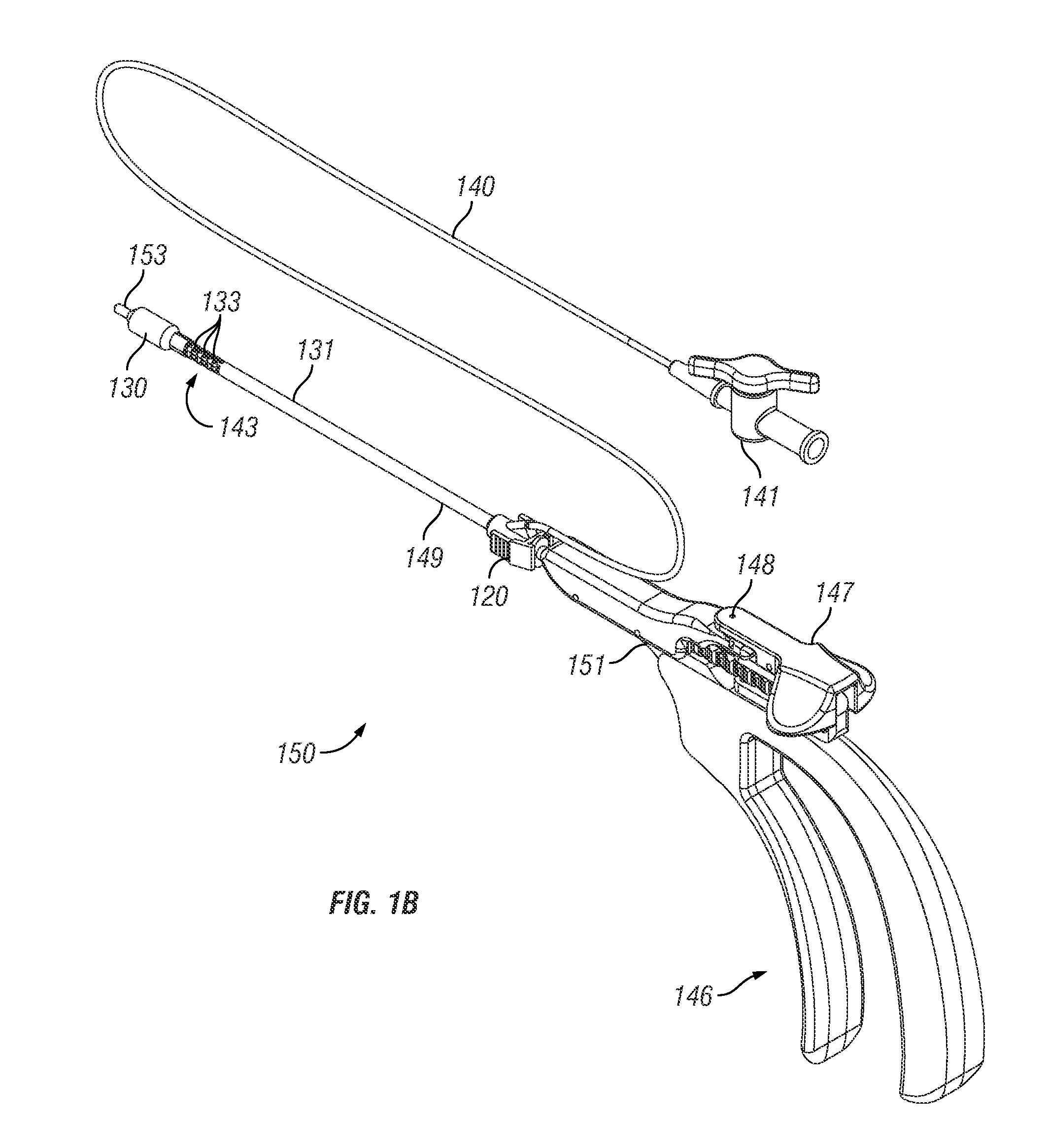
[0019]In an exemplary embodiment, the therapeutic component will interface with a rigid or articulating insertion device. Once interfaced, the device can be easily guided into a desired location using standard surgical techniques, and without the need of other means to guide the device such as guidewires or rigid guide tubes. The handle of the insertion device can include an actuator for controlling the articulation, which will enable the therapeutic component to be positioned and articulated with one hand, leaving the second hand free for holding an endoscope as is standard for FESS surgery. The instrument can also have means for locking the articulation mechanism into certain positions, such that the instrument is effectively rigid at predetermined angles, giving it the feel of standard ENT surgical instrument and providing the ability to accurately position the tip of the device in three-dimensional space. The insertion device can also have provisions and features to enable the intra-operative tracking of the instrument tip using currently available navigation systems. Once the device is in place, the desired therapeutic effect (e.g., dilation, stent placement, etc.) can occur.
[0021]In certain embodiments, the therapeutic component and insertion device include coupling means which allows the therapeutic component to be removably attached to the insertion device, thereby making the therapeutic component interchangeable between different insertion devices during a single procedure. For example, the user may use a single therapeutic component coupled with a variety of articulating and / or rigid instruments to treat all of the sinuses for a single patient. This feature reduces the number of different devices needed for a single procedure, bringing down the cost of the procedure. In an embodiment, the coupling means is attached to an actuator for locking and unlocking the therapeutic component on to the shaft.
[0022]Additional embodiments include features on the insertion device which provide the ability to flush and or suction the ostia, or delivery therapeutic agents, using the same insertion device that delivers the therapeutic component. In addition, embodiments and methods are provided which allow use of a flexible scope to aid in placement of the therapeutic component.
[0032]In certain embodiments, the articulating segments can include a plurality of independent pivot members and pins in an alternating configuration. In particular embodiments, each pivot member can define an opening while each pin can define a pin aperture. In specific embodiments, a first slat assembly and second slat assembly extend through the articulating segments. In certain embodiments, each of the first slat assembly and the second slat assembly is configured to push when the other of the first slat assembly and the second slat assembly pulls so as to cause the articulating segments to articulate.
[0043]In certain embodiments, a bioabsorbable stent may be preferred to reduce the need for removal of the stent once the therapeutic effect has taken place, such as creating patency in the sinus opening throughout the healing period. In another embodiment, the stent may elude medications to create the therapeutic effect. These medications could include anti-inflammatory, antibiotic, steroid, etc. Since typical bioabsorbable stents are rigid, the stent could be composed of multiple leaflets that overlap in a slide and lock design to retain the shape of the ostium once inflated. Alternatively the stent could be composed of a magnesium based alloy that can retain its shape once expanded.
[0084]Particular embodiments comprise a method of dilating a paranasal sinus, where the method comprising: providing a therapeutic component and a flexible endoscope; coupling the therapeutic component to the flexible endoscope; inserting the therapeutic component into a paranasal sinus; utilizing the flexible endoscope to visualize a location within the paranasal sinus; and utilizing the therapeutic component to dilate the paranasal sinus. Certain embodiments comprise providing a light on the flexible endoscope and utilizing the light to transilluminate the sinus. Specific embodiments further comprise using a light on the flexible endoscope to assist in placement of the therapeutic component within the nasal sinus. Certain embodiments further comprise providing an insertion device and coupling the therapeutic component and flexible endoscope to the insertion device to position the therapeutic component in the paranasal sinus. In specific embodiments, the insertion device is articulating, and the method further comprises articulating the delivery device during positioning of the therapeutic component is in the nasal sinus. Particular embodiments further comprise preparing the paranasal sinus to receive the therapeutic component prior to inserting the therapeutic component into the nasal passageway.

Problems solved by technology

Often patients complain of the post-operative pain and bleeding associated with the procedure.
Since FESS is considered an option only for the most severe cases (those showing abnormalities under CT scan), a large population of patients exist that either cannot tolerate the prescribed medications or are not considered candidates for surgery.
To date, there is no mechanical therapy offered for these patients, and even though they may fail pharmaceutical therapies, no other course of action is indicated.
This leaves a large population of patients in need of relief, unwilling or afraid to take steroids, but not sick enough to qualify for surgery.
Also, there is a degree of individual variation in the intranasal and paranasal anatomy of human beings, thus making it difficult to design a stiff-shaft balloon catheter that is optimally shaped for use in all individuals.
Indeed, rigid catheters formed of hypotubes that have pre-set angles cannot be easily adjusted by the physician to different shapes to account for individual variations in the anatomy.
The requirement to test multiple disposable catheters for fit is likely to be very expensive and impractical.
Moreover, if such catheter are disposable items (e.g., not sterilizable and reusable) the need to test and discard a number of catheters before finding one that has the ideal bend angle could be rather expensive.
Furthermore, the rigidity of the catheters described by Becker may make access to certain acutely angled ostia difficult in the confined space of the nasal cavity.
A further disadvantage of Becker is the inability to verify that the balloon position is in the correct location.
In some anatomy where direct visualization is difficult to impossible, for example in the frontal recess, there is a risk of entering and dilating the wrong opening, which at best does not resolve the clinical symptoms and in some cases may lead to severe clinical complications.
However, several disadvantages remain with this approach.
The addition of devices such as guide wires and guide catheters to navigate and position the balloon adds significant complexity and cost to the surgical case.
As described, this added cost and complexity often prohibits these prior systems to be used in conjunction with standard sinus surgery equipment and techniques, but instead be used as a stand-alone procedure for isolated disease.
This factor limits the clinical utility of this prior system, for example it does not allow the concurrent removal of the uncinate process or removal of the ethmoid air cells.
In addition, the techniques employed to use these prior systems are not standard to the average ENT surgeon and require extensive training.
Use of the fluoroscopy system alone requires extensive and expensive additions to operating room equipment, user training, and in some cases user certification.
In addition, as with the Becker system, the guide catheters are shaped with a set angle, so that access to multiple sinuses in one patient may involve the use of several devices, increasing the cost of the procedure still further.
Another disadvantage with the method used to place the balloon catheter, requiring the manipulation of a guide catheter and guide wire, is that this method requires at least two hands, and sometimes a third via an assistant, thus the concurrent use of an endoscope for direct visualization, as is standard for current sinus surgical procedures, would require an assistant: further cost and personnel in the operating room.
The structure of these devices also presents disadvantages.
Because of the lack of rigidity of the guidewire and guide catheter, it is impossible to precisely locate the tips of these devices in 3-D space.
While this is not an issue for vascular procedures where the working space is essentially linear, this is not true for the sinus cavities.
Further, the lack of rigidity of the devices also lessens the ability to push the balloon across the tight spaces often encountered in chronic sinusitis patients, which may be obstructed by scar or granulation tissue.
Finally, the lack of rigidity precludes the use of image guidance navigation systems for positioning and verifying the location of the balloon.
Without removing the uncinate process and diseased tissue of the ethmoid air cells, the potential for surgical failure and need for revision dramatically increases.
Additionally, maintenance of patency of the maxillary, frontal and sphenoid sinus can not be assured by purely balloon dilating the opening, and may require stenting the dilated sinus with an expandable stent to assure patency.
Prior devices, systems and methods have not been optimized for minimally invasive treatment of sinusitis, mucocysts, tumors, infections, hearing disorders, fractures, choanal atresia or other conditions of the paranasal sinuses, Eustachian tubes, Lachrymal ducts and other ear, nose, throat or mouth structures in which the atraumatic dilation and maintance of these structures is desirable.
Non-articulating instruments are not capable of navigating the tortuous pathway to some of these structures.
Guidewire and guide catheter access to these structures may not be possible without risk of trauma to the anatomy, or in some cases may not be possible at all.
Furthermore, these features may also be strong and somewhat elastic so that they do not easily fracture during the process of implantation.
In particular embodiments, the tissue in the paranasal sinus outflow tract is damaged after exposure to the cryogenic temperature.

Method used


Image

  • Systems, devices and methods for providing therapy to an anatomical structure using high frequency pressure waves and/or cryogenic temperatures
  • Systems, devices and methods for providing therapy to an anatomical structure using high frequency pressure waves and/or cryogenic temperatures
  • Systems, devices and methods for providing therapy to an anatomical structure using high frequency pressure waves and/or cryogenic temperatures

Examples


Embodiment Construction

[0155]Exemplary embodiments of the present disclosure provide systems, devices and methods for providing therapy to anatomical structures. In particular embodiments, the therapy comprises dilation of a paranasal sinus. Exemplary embodiments provide the ability to articulate an instrument and maintain the instrument in the articulated position when it is subjected to external forces. This rigidity of the articulated instrument can allow a user to extend the instrument into a paranasal ostium that may include granulation or scar tissue.

[0156]Multiple exemplary embodiments are disclosed in the description that follows. It is understood that various components of the disclosed embodiments can be combined to form additional exemplary embodiments. For example, a handle portion from one disclosed embodiment may be combined with a shaft portion of another disclosed embodiment. Such combinations are within the scope of this disclosure, which is not limited to the specific combinations of fea...


PUM


Abstract

A system, device and method for dilating an anatomical structure. Systems, devices and methods may comprise a therapeutic component configured to treat a paranasal sinus. Specific embodiments may use high frequency pressure waves and / or cryogenic temperatures.

Description

CROSS-REFERENCE TO RELATED APPLICATIONS[0001]This application claims priority to U.S. Provisional Patent Application Ser. Nos. 61 / 386,336 (filed Sep. 24, 2010) and 61 / 387,314 (filed Sep. 28, 2010), the contents of which are incorporated herein by reference.[0002]This application incorporates by reference the following applications: U.S. Provisional Patent Application Ser. Nos. 61 / 184,614 (filed Jun. 5, 2009); 61 / 231,086 (filed Aug. 4, 2009); 61 / 289,480 (filed Dec. 23, 2009); and U.S. patent application Ser. No. 12 / 794,321 (filed Jun. 4, 2010).BACKGROUND OF THE INVENTION[0003]Surgical treatments for ear, nose and throat (ENT) disorders (e.g. sinusitus) have evolved slowly. In current clinical practice, functional endoscopic sinus surgery (FESS) is used to treat disorders where mucous drainage is impaired and / or chronic infections are present. In FESS, an endoscope is inserted into the nose and, under visualization through the endoscope, the surgeon may remove diseased or hypertrophic...

Claims


Application Information

Patent Timeline
Patent Type & Authority Patents(United States)
IPC IPC(8): A61F2/04A61B18/02A61N7/00A61M29/02A61B18/14A61M31/00A61B18/00
CPCA61N7/00A61B18/02A61M29/02A61B18/1492A61B34/20A61B2018/00327A61M31/00A61M2210/0681A61N2007/0043A61N7/022A61B17/24
Inventor GONZALES, DONALD A.DINGER, III, FRED B.NALLURI, PRASAD
Owner ARTHROCARE
Learn More