Shape tunable plasmonic nanoparticles

a plasmonic nanoparticle, shape-tunable technology, applied in the direction of peptides, instruments, therapies, etc., can solve the problems of inconsistent light penetration in tumors, human toxicity and cytotoxicity of semiconductor materials, and risk of severe burns over the patient's entire body, and achieve the effect of quick conversion of light into hea

Active Publication Date: 2017-03-07
RGT UNIV OF CALIFORNIA +1
View PDF3 Cites 0 Cited by
  • Summary
  • Abstract
  • Description
  • Claims
  • Application Information

AI Technical Summary

Problems solved by technology

But the potential human toxicity and cytotoxicity of the semiconductor material is one major problem for its in vivo application.
However, these agents are only relatively selective and risk of severe burns over the patient's entire body when exposed to light persists for 1 to 30 days depending on the agent used.
Further, consistent light penetration in a tumor can be problematic indicating the need for even better photosensitizers and delivery sources.
They all have a common limitation that the heating is excessive and nonspecific and destroys both the malignant and benign cells.
Nanoshells are disclosed in U.S. Pat. No. 6,530,944 The '944 patent discloses that a serious practical limitation to realizing many applications of solid metal nanoparticles is the inability to position the plasmon resonance at technologically important wavelengths, i.e., near infrared wavelengths.

Method used


Image

  • Shape tunable plasmonic nanoparticles
  • Shape tunable plasmonic nanoparticles
  • Shape tunable plasmonic nanoparticles

Examples


example 1

Antibody Conjugated Nanospheres can be Used to Distinguish Between Malignant and Non-Malignant Cells

[0108]A very simple and inexpensive conventional microscope with proper rearrangement of the illumination system and the light collection system was used to imaging cells which were incubated with colloidal gold or with anti-epidermal growth factor receptor (anti-EGFR) antibody conjugated gold nanoparticles. The optical properties of the gold nanoparticles incubated in single living cancerous and noncancerous cells are compared for different incubation methods. It is found that the scattering images and the absorption spectra recorded from anti-EGFR antibody conjugated gold nanospheres incubated with cancerous and noncancerous cells are very different and offer potential techniques for cancer diagnostics.

[0109]One nonmalignant epithelial cell line HaCaT (human keratinocytes), and two malignant epithelial cell lines HOC 313 clone 8 and HSC 3 (human oral squamous cell carcinoma) were cu...

example 2

Light Scattering Images of Antibody Conjugated Nanospheres

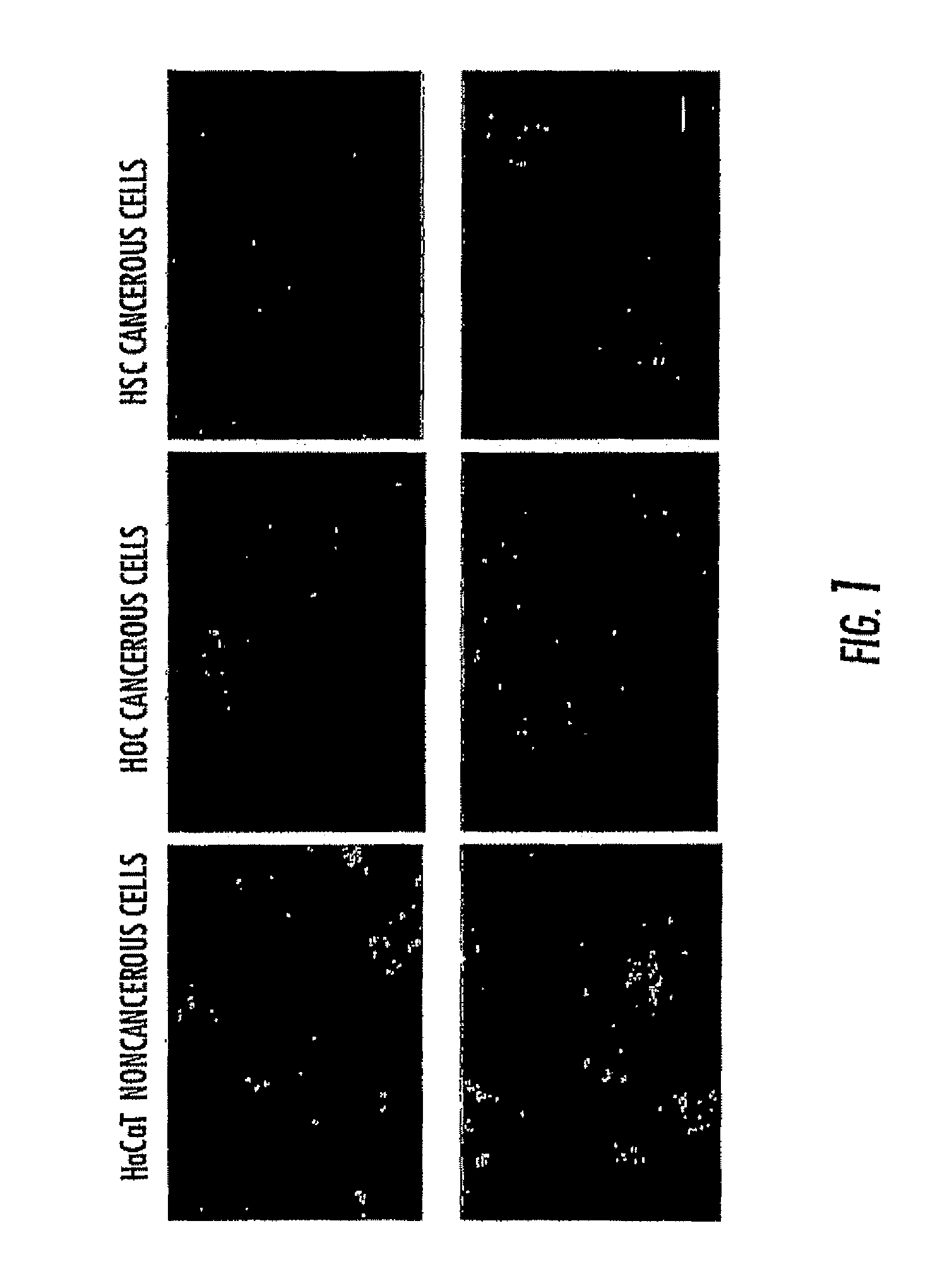
[0115]The light scattering pattern of gold nanoparticles is significantly different when anti-EGFR antibodies were conjugated to gold nanoparticles before incubation with the cells (FIG. 3). The HaCaT on cancerous cells are poorly labeled by the nanoparticles and the cells could not be identified individually (FIG. 3, three images on the left column). When the conjugates are incubated with HOC FIG. 3, three images on the middle column) and HSC (FIG. 3, tree images on the right column) cancerous cells for the same amount of time, the nanoparticles are found on the surface of the cells, especially on the cytoplasm membranes for HSC cancer cells. This contrast difference is due to the specific binding of overexpressed EGFR on the cancer cells with the anti-EGFR antibodies on the gold surface. The nanoparticles are also found on the HaCaT noncancerous cells due to part of the specific binding, but mostly due to the nonspecific in...

example 3

Selectivity of Anti-EGFR Antibody Conjugated Au Nanoparticles

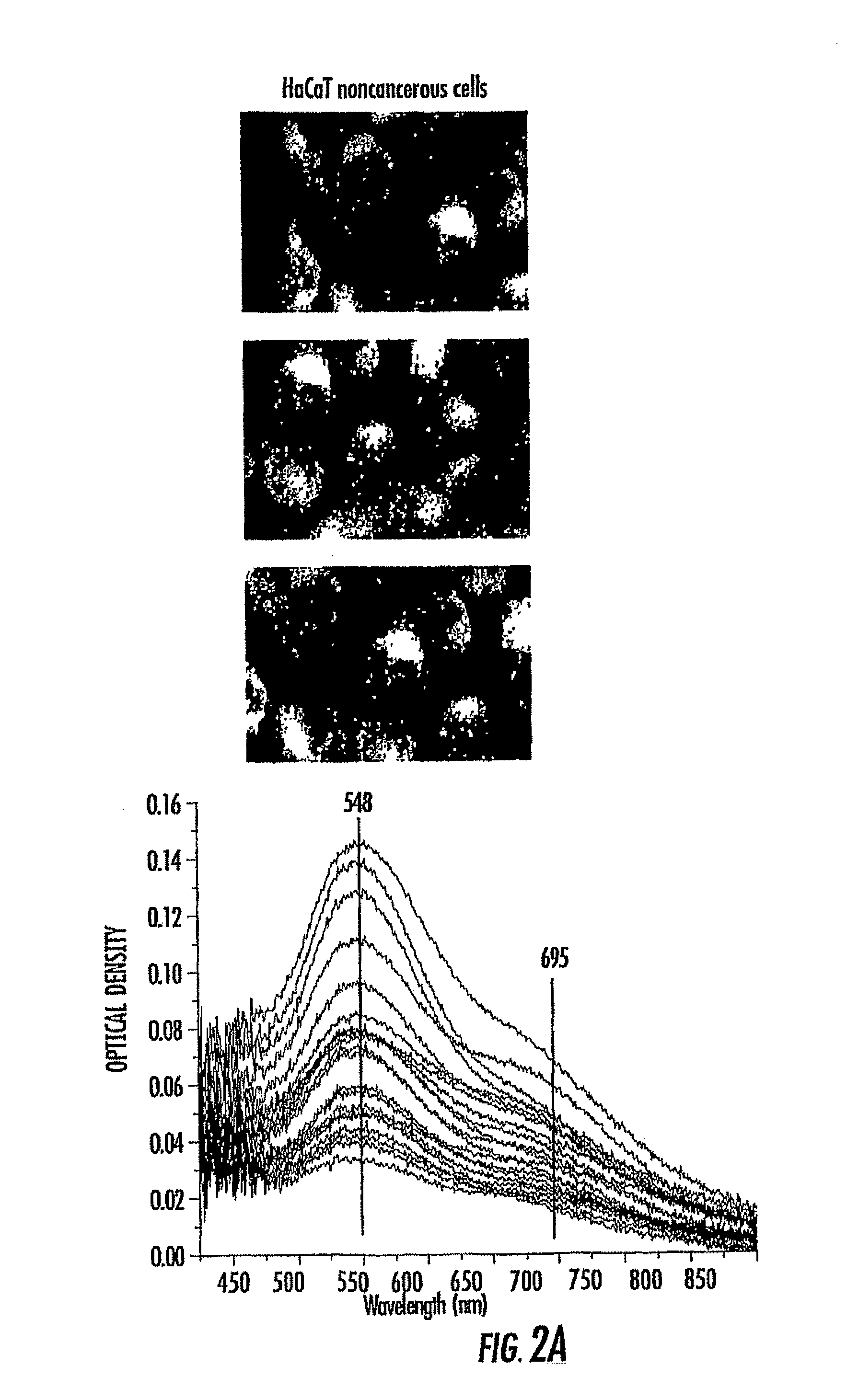
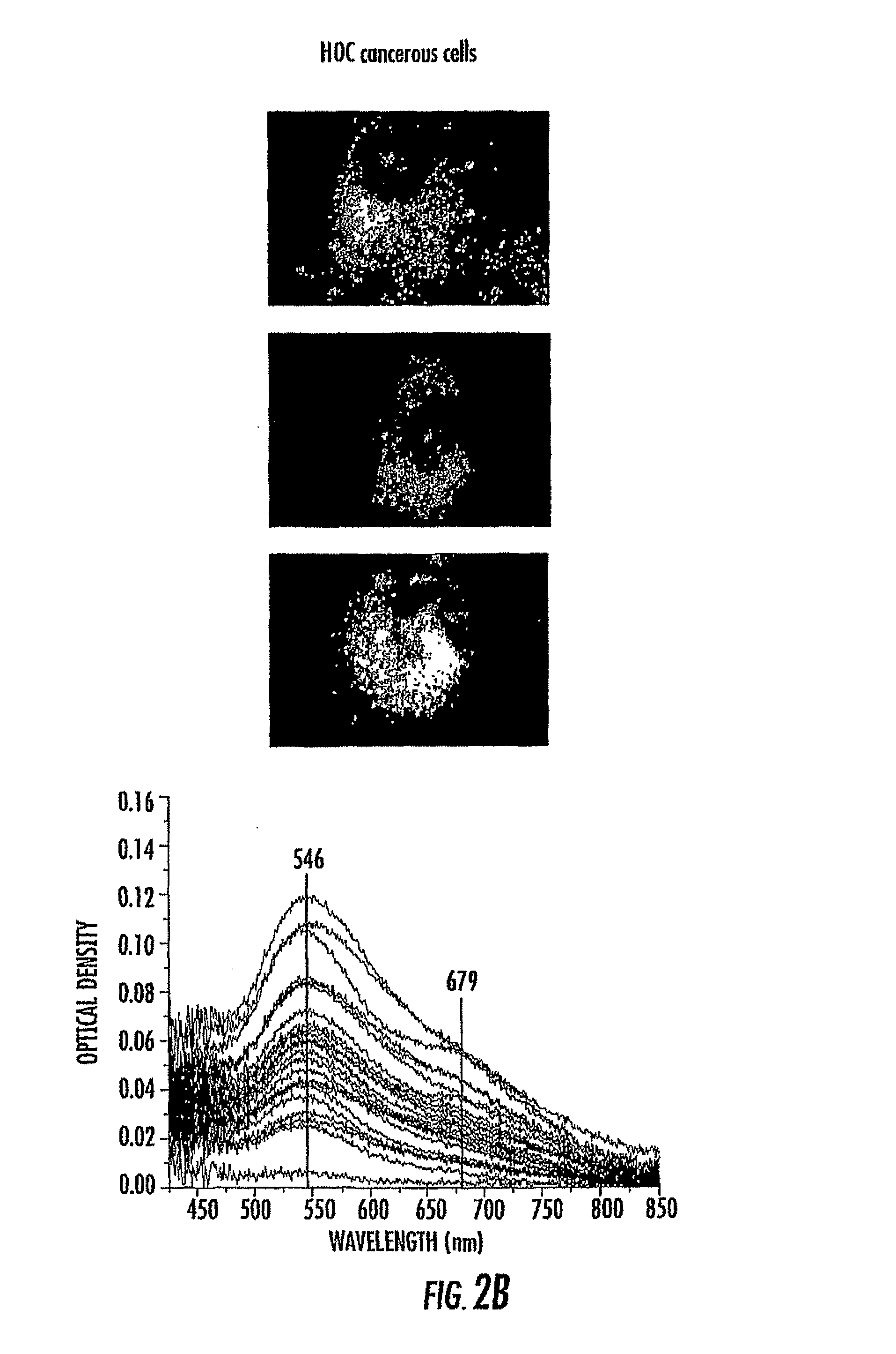
[0118]The strongly scattered colored light of gold nanoparticles is easily observed in a simple dark field setup. In live cell cultures, immuno-targeted nanoparticles successfully label and distinguish the malignant HOC and HSC cells from the HaCaT benign cells. The surface plasmon absorption spectrum itself can differentiate between the malignant and benign cells. FIG. 4 shows the light scattering pattern of anti-EGFR antibody conjugated gold nanoparticles on the surface of benign and malignant cells that are used in the laser irradiation experiment. The HaCaT cells (FIG. 4, left) are poorly labeled with the nanoparticles. When the Au conjugates are incubated with USC (FIG. 4, middle) and HOC (FIG. 4, right) for an equal amount of time, the cell surfaces are heavily labeled with the gold nanoparticles due to the specific binding of the anti-EGFR antibodies to the overexpressed EGFR on the surface of the carcinoma cells. T...


PUM

PropertyMeasurementUnit
aspect ratioaaaaaaaaaa
wavelengthaaaaaaaaaa
lengthaaaaaaaaaa

Abstract

Noble metal nanoparticles and methods of their use are provided. Certain aspects provided solid noble metal nanoparticles tuned to the near infrared. The disclosed nanoparticles can be used in molecular imaging, diagnosis, and treatment. Methods for imaging cells are also provided.

Description

CROSS REFERENCE TO RELATED APPLICATION[0001]This application is a filing under 35 U.S.C. §371 of PCT / US2006 / 018177 filed with the U.S. Receiving Office of the Patent Cooperation Treaty on May 11, 2006, which claims priority to U.S. Provisional Patent Application No. 60 / 679,901 filed on May 11, 2005, and U.S. Provisional Patent Application No. 60 / 719,360 filed on Sep. 22, 2005, and U.S. Provisional Patent Application No. 60 / 792,016 filed on Apr. 16, 2006, and where permissible, all of which are incorporated by reference in their entirety.STATEMENT REGARDING FEDERALLY SPONSORED RESEARCH OR DEVELOPMENT[0002]Aspects of the invention were funded in part by Grant Number DE-FG02-97ER14799 awarded by the Department of Energy. The U.S. Government has certain rights in the invention.BACKGROUND[0003]1. Technical Field[0004]Aspects of the disclosure are generally directed to compositions comprising noble metal plasmonic nanoparticles (those that have characteristic strong surface plasmon resona...

Claims


Application Information

Patent Timeline
Patent Type & Authority Patents(United States)
IPC IPC(8): G01N33/58B82Y5/00A61K41/00A61K49/00
CPCG01N33/587A61K41/0052A61K49/0065B82Y5/00B22F2998/00B22F1/0018B22F1/0025B22F1/0547B22F1/054
Inventor EL-SAYED, MOSTAFA A.EL-SAYED, IVAN HOMER
Owner RGT UNIV OF CALIFORNIA
Why Eureka
Try Eureka