Cell culture
a cell culture and cell technology, applied in the field of cell culture, can solve the problems that the single-cell micro-well technology is not suitable for high-resolution imaging
- Summary
- Abstract
- Description
- Claims
- Application Information
AI Technical Summary
Benefits of technology
Problems solved by technology
Method used
Image
Examples
example 2
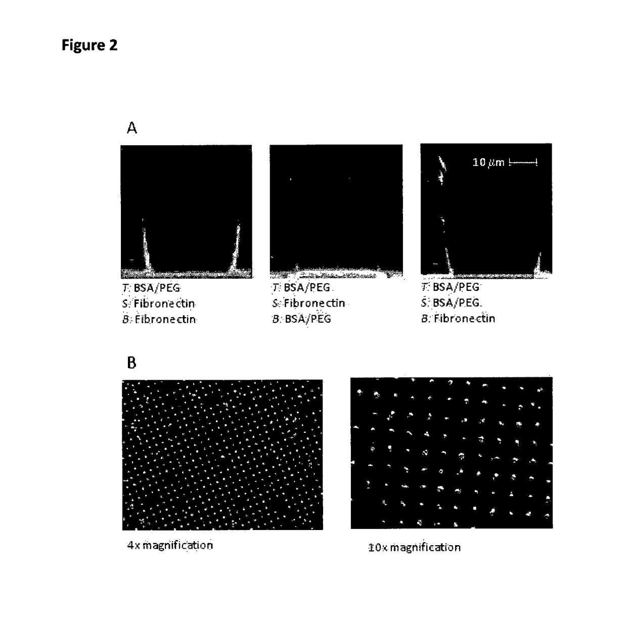
[0127]FIG. 2 illustrates the various combinations we used. Various other combinations where tested including BSA, cadherins and collagen. Though they are possible they are not used for hepatocyte cultures. The protein coating is mainly due to protein adsorption. Using a heterobifunctional PEG molecule Acrylate-PEG-NHS from NANOCS (nanocs.com), we are currently investigating the possibility to perform a direct chemical grafting. The acrylate moiety reacts with the polymer under UV curing and the NHS moiety is a standard functional group for protein functionalization.
example 3
[0128]Primary hepatocytes freshly obtained from rat liver according to [22], are seeded at a density of 0.5 million / mL onto the Petri dish. Protecting the top of the membrane with an antifouling treatment is a key component to help cells fall in the wells. Failure in the antifouling treatment will result in cells attaching to the membrane top and not falling in the microwells. The seeding is followed by a gentle shaking procedure described in protocol to ensure the maximum well filling capability. Following this protocol 90% of the wells are filled with at least one cell and 70% are filled with at least two. Better optimization of the doublet rate is under current investigation.
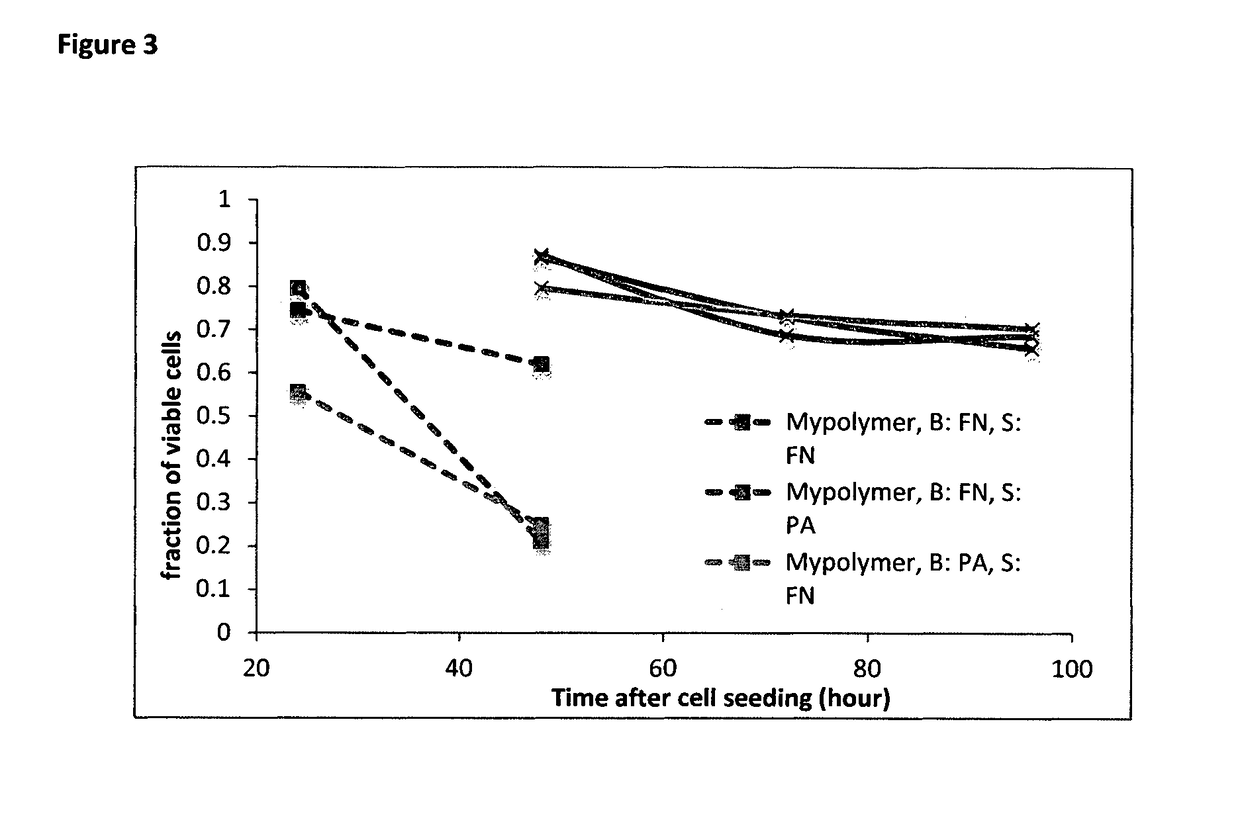
[0129]The viability of the cells was tested daily since 24 h after cell seeding with propidium iodine solution (Sigma). In microwells made by MyPolymer, cells in more than 50% of filled wells are viable at 24 h after seeding even though the survival rate drops to about 20% at 48 h after seeding. However, for ...
example 4
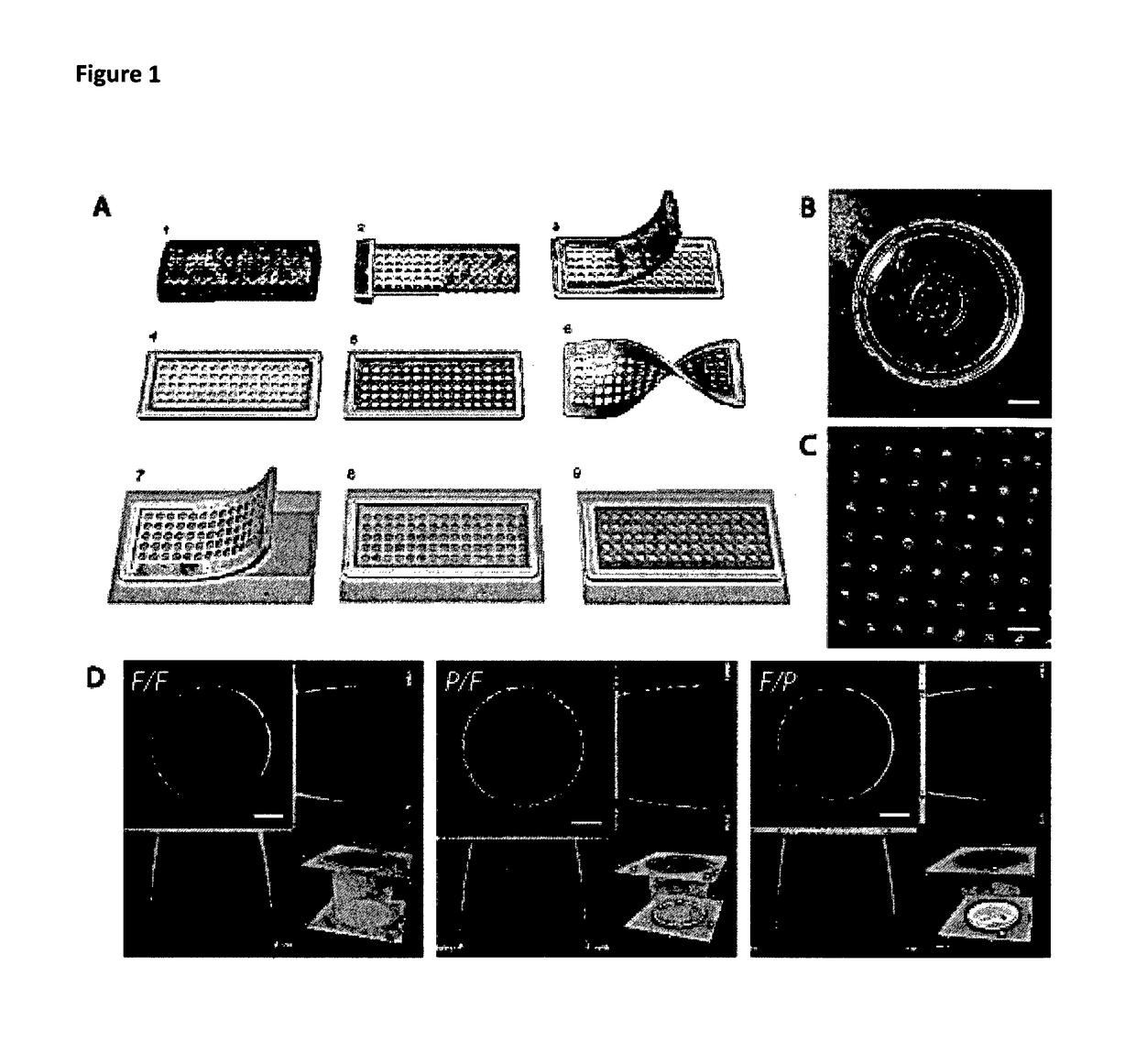
[0130]The hepatocytes can be fixed and stained with antibody within the wells. High resolution imaging within a single well without loss of optical resolution compared to normal culture conditions can then be performed. Live imaging with transfected protein is also possible. This offers the opportunity to monitor the evolution of a series of single doublets over time in a very easy way. As shown in FIG. 4 the morphology and orientation of the bile canaliculi can be tuned by changing the chemical coating of the wells. That property can be used to control the tension on the bile canaliculi and its correlation with drug testing. The formation of vertical canaliculi also allows high spatial and time resolution imaging of molecular transport from the cytoplasm to the bile duct. It is a first step towards the control of the formation of bile canaliculi spanning several cells. In addition the formation of vertical bile canaliculi makes it handier to collect bile secretion which is a key ch...
PUM
| Property | Measurement | Unit |
|---|---|---|
| diameter | aaaaa | aaaaa |
| aspect ratio | aaaaa | aaaaa |
| refractive index | aaaaa | aaaaa |
Abstract
Description
Claims
Application Information
 - R&D
- Intellectual Property
- Life Sciences
- Materials
- Tech Scout
- Unparalleled Data Quality
- Higher Quality Content
- 60% Fewer Hallucinations
Browse by: Latest US Patents, China's latest patents, Technical Efficacy Thesaurus, Application Domain, Technology Topic, Popular Technical Reports.
© 2025 PatSnap. All rights reserved.Legal|Privacy policy|Modern Slavery Act Transparency Statement|Sitemap|About US| Contact US: help@patsnap.com


