Tissue scaffold materials for tissue regeneration and methods of making

a tissue scaffold and tissue technology, applied in the direction of prosthesis, bandages, pharmaceutical delivery mechanisms, etc., can solve the problems of high failure rate of tissue scaffolds in complex settings, avascularity of tissue scaffolds, and insufficient neovascularization of existing tissue replacement products, and achieve the effect of increasing density

Active Publication Date: 2018-05-15
CORNELL UNIVERSITY
View PDF7 Cites 3 Cited by
  • Summary
  • Abstract
  • Description
  • Claims
  • Application Information

AI Technical Summary

Benefits of technology

[0008]Disclosed herein is a type of tissue scaffold material made of a hydrogel with embedded microspheres. In the disclosed tissue scaffolds, the microspheres have a different or greater density (w / v) of polymer relative to the density of the hydrogel, which differential density facilitates cellular invasion into the tissue scaffold. In one embodiment, the hydrogel includes a first polymer and the microspheres include a second polymer, the microspheres are embedded in the hydrogel, and the microspheres have a greater density than the hydrogel.

Problems solved by technology

The use of currently available tissue replacement scaffolds is not without substantial associated cost.
In addition, commercially available tissue scaffolds are avascular and prone to high failure rates when used in complex settings, such as irradiated wounds or those with exposed hardware or bone.
In such complex settings, neovascularization is insufficient using existing tissue replacement products.

Method used


Image

  • Tissue scaffold materials for tissue regeneration and methods of making
  • Tissue scaffold materials for tissue regeneration and methods of making
  • Tissue scaffold materials for tissue regeneration and methods of making

Examples


example 1

n of Microsphere / Hydrogel Scaffolds

[0055]Collagen type I was extracted from rat tail samples using standard techniques. Skin was removed from rat tails using sharp dissection and discarded. Then, starting from the distal end of the tail, tendons were extracted by breaking a joint within the vertebrae and pulling upward on the distal vertebrae until the distal vertebrae with attached tendon separated from the remaining proximal tail. The vertebrae was then sharply dissected from the tendon and discarded. Next, the tendon was placed in 70% ethanol. This was repeated until all joints within the tail were broken and tendons extracted. The extracted tendons were collected, weighed and placed in a sterile 1 L container. Thereafter, 0.1% acetic acid was added to the tendons to reach a final concentration of 75 ml of acetic acid / g of tendon in order to arrive at a stock collagen solution of 15 mg / mL (1.5% w / v) type I collagen. The collagen stock was then stored at 4 C and agitated for appro...

example 2

re Containing Scaffolds Promote Cellular Infiltration

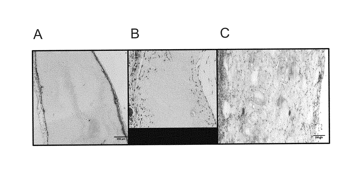
[0062]Scaffolds were produced one day prior to implantation. Scaffolds were implanted subcutaneously in the dorsa of 8 week old wild-type C57bl / 6 mice. 3 mice were implanted with 4 total scaffolds as follows: Two 1% microspheres in 0.3% bulk scaffolds; one 1% bulk scaffold as a control; one 0.3% bulk scaffold as a control. All mice were sacrificed and harvested for histological analysis after 7 or 14 days. Hematoxylin and eosin (H&E) staining was performed on tissue samples embedded in optimal cutting temperature compound (OCT) medium, to identify cellular infiltration into scaffolds.

[0063]After 7 days of implantation, the microsphere scaffolds (MSS) show substantial and uniform cellular invasion spanning the entire depth of the scaffold (FIG. 1C). Comparatively, cells sporadically and only partially invaded the 0.3% control scaffolds (FIG. 1B), and failed to invade the 1% control scaffolds, instead proliferating along the periphe...

example 3

Densities of Microspheres Relative to Hydrogel Density Promote Cellular Infiltration

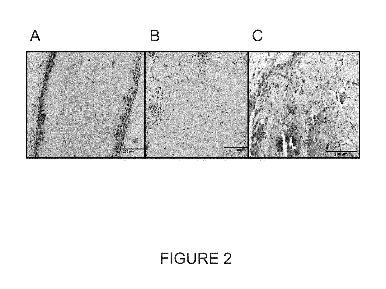
[0065]Microsphere scaffolds with different densities (w / v) of collagen in microsphere (MS) and hydrogel (H) were prepared as follows: (A) 1% collagen MS in 0.3% H; (B) 0.6% MS / 0.3% H; (C) 0.4% MS / 0.2% H; (D) 0.4% MS / 0.6% H. See, Table 1.

[0066]

TABLE 1Densities of Microsphere ScaffoldsMicrosphere Collagen Density (w / v)Bulk Collagen Density (w / v) 1%0.3%0.6%0.3%0.4%0.2%0.4%0.6%

[0067]MSS were implanted subcutaneously in the dorsa of adult mice and harvested for immunohistochemistry at 7 and 14 days after implantation. Immunohistochemical analysis identified cellular infiltration in all MSS (FIGS. 3A-3D), with greatest infiltration seen in 1% MS / 0.3% H, and 0.6% MS 0.3% H (FIGS. 3C-3D). In addition, CD31 expression was seen in all MSS after 7 and 14 days of implantation (FIGS. 4A-4B), indicative of invading endothelial precursors and the formation of neovasculature.


PUM

PropertyMeasurementUnit
diameteraaaaaaaaaa
diameteraaaaaaaaaa
diameteraaaaaaaaaa

Abstract

Disclosed herein are tissue scaffold materials with microspheres of one density embedded in hydrogel of a different density. The disclosed materials have improved ability to facilitate cellular invasion and vascularization for wound healing and tissue regeneration. The inventors have found that materials having components with different densities promotes invasion of cells, including desirable cells such as fibroblasts and endothelial precursor cells, into the scaffold.

Description

CROSS-REFERENCE TO RELATED APPLICATIONS[0001]This application claims priority to U.S. provisional application 61 / 906,131, filed Nov. 19, 2013, which is incorporated herein in its entirety.BACKGROUND OF THE DISCLOSURE[0002]The optimization of cell guidance through autologous or artificial tissue scaffolds has long been a topic of great interest. The most prevalent and thus far the most successfully applied off-the-shelf “tissue-engineered” products were all originally intended to serve as dermal replacement scaffolds. Commercially available scaffolds are acellular and thus share the common requirements of host cell invasion and vascularization to achieve durable incorporation. Because this process is prolonged, requiring a minimum of several weeks for completion and necessitating obligatory dressing changes, wound immobilization, and nursing care, there is significant interest in developing better scaffolds that could optimize the rate of cellular invasion. (Eppley, Plast Reconstr Su...

Claims


Application Information

Patent Timeline
Patent Type & Authority Patents(United States)
IPC IPC(8): A61K9/00A61L27/54A61K9/16A61L27/26A61L27/48A61L27/52A61L27/44A61L26/00A61L27/60
CPCA61L27/48A61K9/0024A61K9/16A61K9/1635A61K9/1647A61L26/008A61L26/0052A61L27/26A61L27/44A61L27/52A61L27/54A61L27/60C08L89/06A61L2300/602A61L2300/412A61L27/24A61L27/56A61L2430/34
Inventor SPECTOR, JASONSTROOCK, ABRAHAM D.MORGAN, JOHN
Owner CORNELL UNIVERSITY
Why Eureka
Try Eureka