Acellular dermal matrix and its preparing method

A decellularized dermis and matrix technology, applied in skin transplantation, medical science, prosthetics, etc., can solve the problems of limited inactivation methods for mad cow disease pathogens, the risk of spreading viruses, and narrow sources of raw materials, etc., to achieve high practical application value and source Wide range of effects with good tissue compatibility

Active Publication Date: 2008-03-05
YANTAI ZHENGHAI BIO TECH
View PDF10 Cites 2 Cited by
  • Summary
  • Abstract
  • Description
  • Claims
  • Application Information

AI Technical Summary

Problems solved by technology

But at the same time, there are some shortcomings: 1. The source of raw materials is narrow, there are ethical concerns, and it is not easy to popularize; 2. The price is expensive; 3. There is a risk of spreading viruses.
Currently, methods for inactivating the BSE pathogen are limited

Method used


Image

  • Acellular dermal matrix and its preparing method
  • Acellular dermal matrix and its preparing method
  • Acellular dermal matrix and its preparing method

Examples


Embodiment 1

[0043] Embodiment 1, the preparation of acellular dermal matrix

[0044] Using sheep as the material source for preparing the acellular dermal matrix, the specific preparation process includes the following steps:

[0045] 1) peeling off the body surface skin of the sheep;

[0046] 2) Remove subcutaneous fat, muscle and hair and other incidental components;

[0047] 3) Wash the skin with clean water;

[0048] 4) Treat the skin with 2N sodium hydroxide solution for 40 minutes;

[0049] 5) replace with new 2N sodium hydroxide solution, and then treat for 40 minutes;

[0050] 6) Step 5) is repeated once;

[0051] 7) Treat the skin with 5% Tween-80 for 20 minutes;

[0052] 8) The skin treated by the above steps is fully washed with depyrogenated water to remove the residual chemical components to obtain the acellular dermal matrix, which is stored in PBS.

Embodiment 2

[0053] Embodiment 2, preparation of decellularized dermal matrix

[0054] Using cattle as the material source for preparing the acellular dermal matrix, the specific preparation process includes the following steps:

[0055] 1) peeling off the body surface skin of the cow;

[0056] 2) Remove subcutaneous fat, muscle and hair and other incidental components;

[0057] 3) Wash the skin with clean water;

[0058] 4) Treat the skin with 5N sodium hydroxide solution for 30 minutes;

[0059] 5) replace with new 5N sodium hydroxide solution, and then treat for 30 minutes;

[0060] 6) Step 5) is repeated twice;

[0061] 7) Treat the skin with 1% TRITON-X100 for 30 minutes;

[0062] 8) The skin treated by the above steps is fully washed with depyrogenated water to remove the residual chemical components to obtain the acellular dermal matrix, which is stored in PBS.

Embodiment 3

[0063] Example 3, Structural Observation and Detection of Acellular Dermal Matrix

[0064] 1. Observation of electron microscope structure of acellular dermal matrix

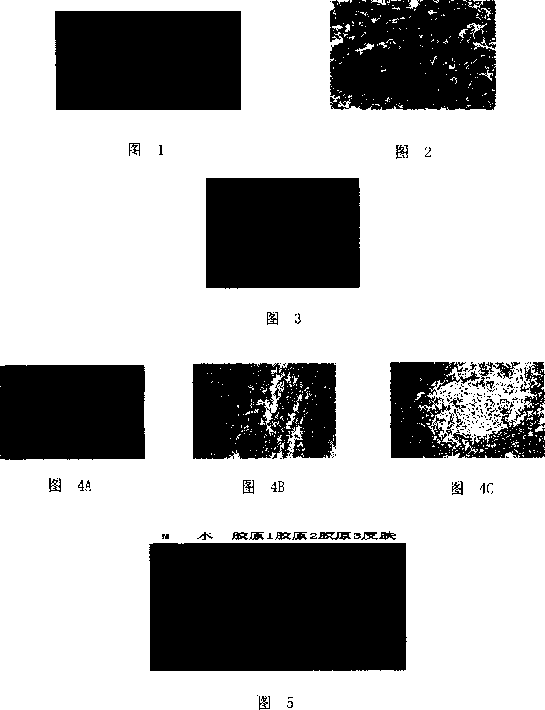
[0065] The freeze-dried photos of the acellular dermal matrix prepared in Example 1 and Example 2 are shown in Figure 1, and then its internal structure is observed with an electron microscope, as shown in Figure 2, indicating that the material space maintains a size of 20-100 microns The prepared acellular dermal matrix is ​​suitable for the growth of various types of cells.

[0066] 2. Biocompatibility testing of acellular dermal matrix

[0067] Human fibroblasts were inoculated on the acellular dermal matrix prepared in Example 1 and Example 2, and the number of inoculated cells was 10 6 One was cultured at 37°C, and the growth of human fibroblasts was observed. The growth of the cells after inoculation was shown in Figure 3. The growth of human fibroblasts was vigorous, indicating that the prepared acellul...


PUM


Abstract

The present invention discloses a cell-removed hypodermal matrix and its preparation method. Its preparation method includes the following steps: (1) using skin of mammal body surface as raw material; (2) removing hypodermic incidental components; (3) cleaning skin; (4) treating skin by using 2-5N sodium hydroxide solution for 10-60min; (5) using new 2-5N sodium hydroxide solution to treat skin for 10-60 min; (6) repeating step 5 for 1-3 times; (7) using detergent whose mass percentage concentration is 1-5% to treat said skin treated by sodium hydroxide solution; and (8) cleaning said skin by using hot raw water so as to the invented product.

Description

technical field [0001] The invention relates to an acellular dermal matrix and a preparation method thereof. Background technique [0002] The study of acellular dermal matrix has gone through a long process. As early as 1913, Loewe confirmed that dermal tissue can enhance the toughness of grafted skin, inhibit the growth of granulation tissue and scar formation, and reduce the degree of wound contracture. Tissues play an important role in skin transplantation; in 1985, Heck et al. reported that allogeneic dermis was used as the graft carrier of autologous epidermis, and the two were compositely transplanted on the escharectomy wounds of animal models and burn patients, and achieved good wound repair and scar inhibition. proliferative effect. However, subsequent applications and research have found that the immune rejection of allogeneic skin transplantation is mainly derived from cells such as epidermal cells, endothelial cells, and fibroblasts, thus promoting the study of...

Claims


Application Information

Patent Timeline
Patent Type & Authority Patents(China)
IPC IPC(8): A61F2/10A61L27/60
Inventor 董群林萍孙先昌
Owner YANTAI ZHENGHAI BIO TECH
Why Eureka
Try Eureka