Biological membrane fixing device for eye surface

A fixing device and biofilm technology, applied in the field of medical devices, can solve the problems of fragile perforation, multiple operations, traumatic ocular surface, etc., and achieve the effect of improving the treatment effect, requiring no surgical operation, and simple operation.

Active Publication Date: 2009-11-25
ZHONGSHAN OPHTHALMIC CENT SUN YAT SEN UNIV
View PDF4 Cites 0 Cited by
  • Summary
  • Abstract
  • Description
  • Claims
  • Application Information

AI Technical Summary

Problems solved by technology

In most cases, repeated amniotic membrane coverage can achieve the therapeutic purpose. However, the method of fixing the amniotic membrane in the current clinical application of amniotic membrane coverage is mainly surgical suture, which will inevitably damage the ocular surface. Because the amniotic membrane is thin, it is easy to break and perforate. Multiple surgeries are often required to achieve therapeutic goals

Method used


Image

  • Biological membrane fixing device for eye surface
  • Biological membrane fixing device for eye surface
  • Biological membrane fixing device for eye surface

Examples


Embodiment 1

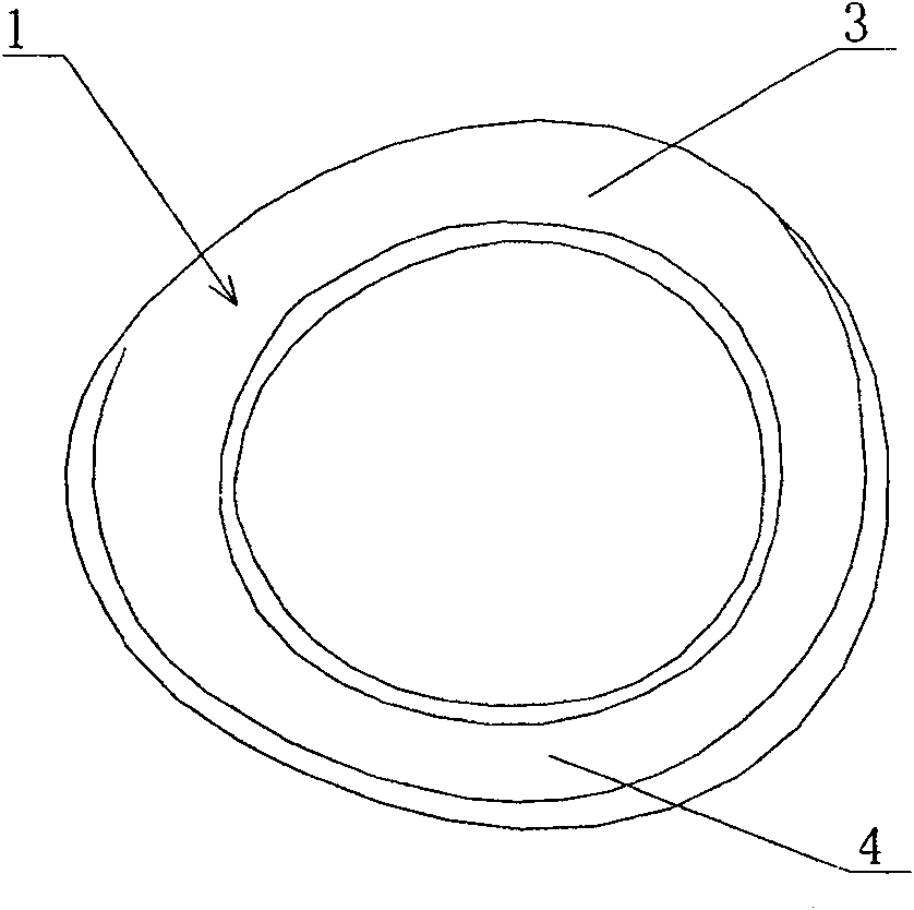
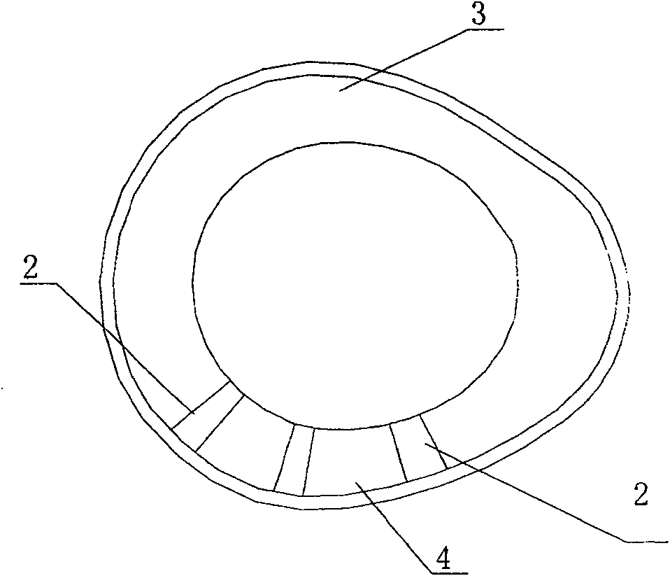
[0025] Such as Figure 1 to Figure 2 As shown, the fixation device 1 of this embodiment has a circular shape with a radian, including an upper fornix 3 and a lower fornix 4, and its radian coincides with the radian of the patient's eyeball surface and the upper and lower fornix conjunctiva. The fixing device is elliptical and is made of polymethyl methacrylate (PMMA). On the inner surface of the fixing device, that is, the side where the fixing device contacts the eyeball, there are three grooves 2 arranged radially. The groove 2 is arranged in the lower fornix.

[0026] The inner surface of the fixing device, that is, the side where the fixing device is in contact with the eyeball, can also be provided with more than 3 grooves arranged radially, such as 5 or 7; it is also possible to set only 1 in the lower fornix according to the specific conditions of the disease. groove 2.

Embodiment 2

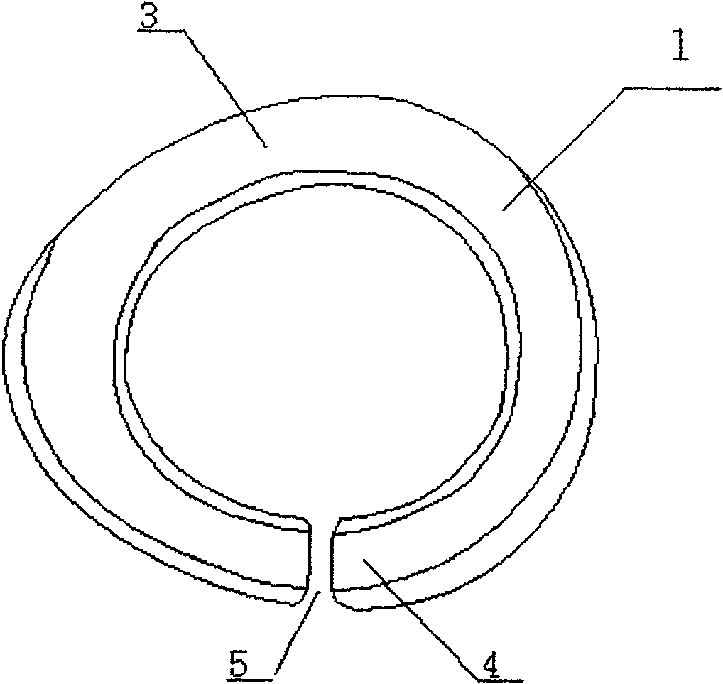
[0028] Such as image 3 As shown, the fixation device 1 in this embodiment is annular with a radian, including an upper fornix 3 and a lower fornix 4, and its radian coincides with the radian of the patient's eyeball surface and the upper and lower fornix conjunctiva. It is elliptical and made of glass. The fixing device is in a non-airtight ring shape, that is, there is a gap 5 at the lowest part of the lower fornix 4 of the fixing device.

Embodiment 3

[0030] Such as Figure 4 or Figure 5 As shown, the fixation device 1 in this embodiment is annular with a radian, including an upper fornix 3 and a lower fornix 4, and its radian coincides with the radian of the patient's eyeball surface and the upper and lower fornix conjunctiva. It is elliptical and is made of silicone gel. The fixing device is a non-airtight ring, that is, there is a gap 5 at the lowest part of the lower fornix 4 of the fixing device. The center has a mesh structure 6, and the mesh structure 6 can adopt such as Figure 4 or Figure 5 shown in the manner.

[0031] The above-mentioned ocular surface biofilm fixation device is used for the treatment of fixed ocular surface defects of patients, and the steps are as follows:

[0032] (1) The patient lies on his back or sits with his head tilted back, and the eyelid skin is disinfected with fluorine iodine.

[0033] (2) According to the size and shape of the patient's conjunctival sac, select a suitable ase...


PUM


Abstract

The invention discloses an ocular surface biofilm fixation device. The fixation device is annular with a radian, and the radian coincides with the radians of the eyeball surface and the upper and lower fornix conjunctiva. The inner surface of the fixation device (in contact with the eyeball surface) can also be provided with grooves. The fixation device for ocular surface biofilm is easy to operate and does not require surgery; the amniotic membrane can be replaced repeatedly at any time to maximize the use of amniotic membrane to resist infection, promote the recovery of injured ocular surface cells, inhibit new blood vessels, and inhibit the activity of collagenase inhibitors ; avoid symblephane and reduce fornix narrowing; avoid subamniotic effusion; do not cause new trauma to the ocular surface, reduce the occurrence and development of corneal limbal granulation, can cover the cornea, bulbar conjunctiva surface, palpebral conjunctiva surface, fornix conjunctiva surface, eliminate It eliminates the dead angle of amniotomy coverage; it does not need to use instruments or sutures to open the eyelids like amnion coverage, so that it does not damage the eyelids and other advantages.

Description

technical field [0001] The invention relates to a medical device, and more particularly relates to a fixation device for an ocular surface biofilm. Background technique [0002] The biomembrane used to treat ocular surface damage is mainly amniotic membrane, and may also be other membranous matter with cells. The amniotic membrane is the part of the fetal membrane in contact with the amniotic fluid, which is differentiated from the trophoblasts, with a thickness of 0.02-0.05 mm, translucent, tough, without blood vessels, nerves, and lymphatic vessels. Divided into five layers: epithelial cell layer, basement membrane, parenchymal layer, fibroblast layer and spongy layer. The epithelium is a single layer of cuboidal cells, which may also be pseudostratified. Under the electron microscope, 0.6 μm microvilli on the surface of the amniotic epithelial cells can be seen. The basement membrane is a cell-free network of fibers. The parenchymal layer is thin and dense, loosely co...

Claims


Application Information

Patent Timeline
Patent Type & Authority Patents(China)
IPC IPC(8): A61F9/00
Inventor 王智崇陈家祺葛坚陈冬梁轩伟
Owner ZHONGSHAN OPHTHALMIC CENT SUN YAT SEN UNIV
Why Eureka
Try Eureka