Integration micro flow control chip used for apoptosis research and application thereof
A microfluidic chip and cell technology, applied in the field of cell apoptosis research, can solve the problems of long processing time, difficulty in real-time analysis of cell apoptosis kinetics, cumbersome operation, etc., and achieve the effect of reducing the consumption of cells and reagents
- Summary
- Abstract
- Description
- Claims
- Application Information
AI Technical Summary
Problems solved by technology
Method used
Image

Examples
Embodiment 1
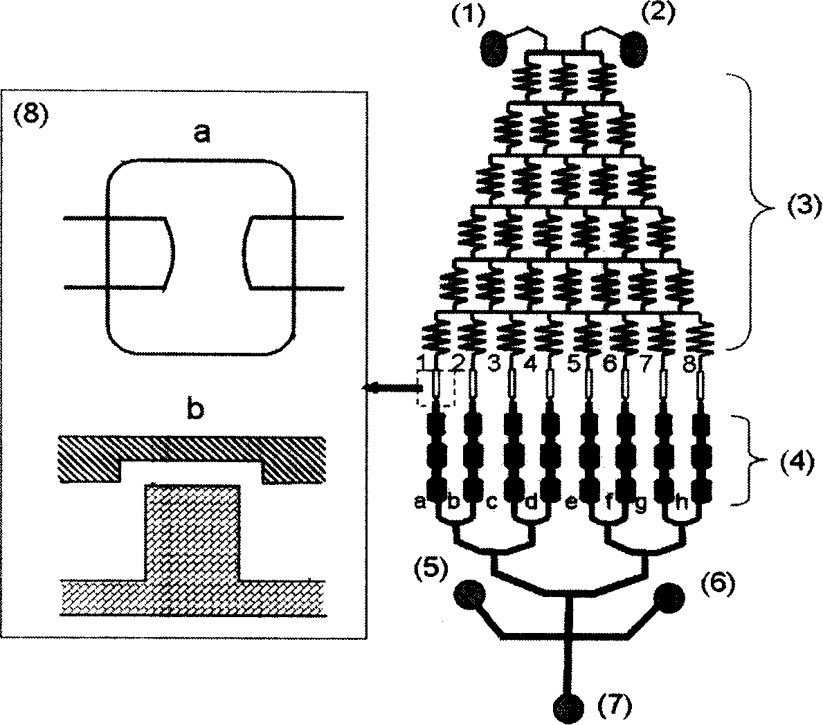
[0034] use figure 1The microfluidic chip shown was used to study the morphological changes of apoptosis. After the chip was inoculated with human hepatocellular carcinoma (HepG2), place it at 37°C in 5% CO 2 6 hours in the incubator. After the cells adhered to the wall, add DMEM medium (high sugar) with adriamycin concentrations of 0 μM and 4.0 μM, place the chip in the cell culture incubator for 24 hours, and keep the solution flowing from the concentration gradient generation unit to the cells at a small flow rate. culture unit. Phosphate buffer saline (PBS) was added to wash the cells, and the chip was observed under an optical microscope. Then in 1mg mL -1 acridine orange Cells were labeled with ethidium bromide and immediately viewed under a fluorescence microscope. Such as figure 2 As shown, doxorubicin induces apoptosis of HepG2 cells, and presents typical morphological changes of apoptosis: cells shrink, become round, and the spacing becomes larger; chromatin ...
Embodiment 2
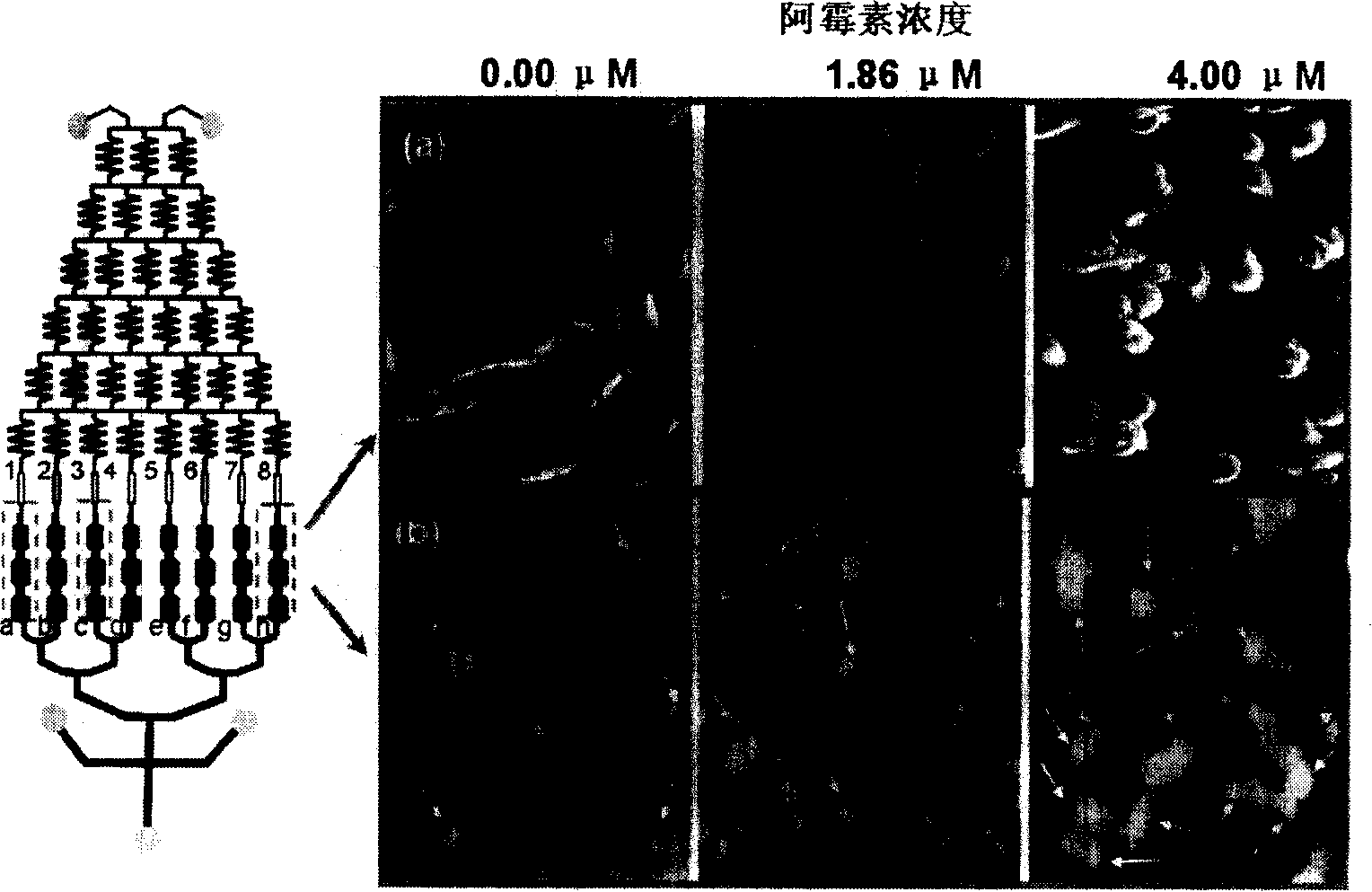
[0036] use figure 1 The microfluidic chip shown was used to study cell membrane changes during cell apoptosis. Chips were inoculated with HepG2 and placed at 37°C, 5% CO 2 6 hours in the incubator. After the cells adhered to the wall, add DMEM medium (high sugar) with adriamycin concentrations of 0 μM and 4.0 μM, place the chip in the cell culture incubator for 24 hours, and keep the solution flowing from the concentration gradient generation unit to the cells at a small flow rate. culture unit. Add PBS solution to wash the cells, add the labeling solution Annexin V / PI, place the chip in the dark for 15 minutes, and finally observe the chip under a fluorescent microscope. As shown in Figure 3, doxorubicin induces characteristic changes in the HepG2 cell membrane: in the early stage of apoptosis, the phosphatidylserine (PS) located on the inner side of the cell membrane is turned outward, showing a green circle (●); in the late stage of apoptosis, the cell membrane is permea...
Embodiment 3
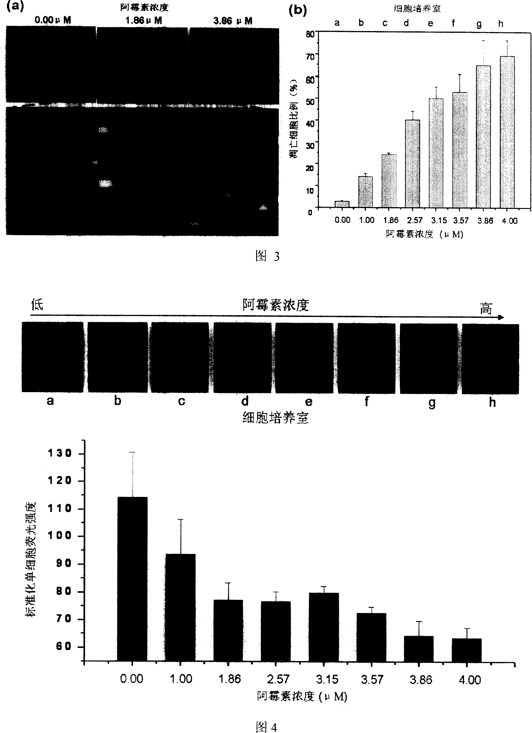
[0038] use figure 1 The microfluidic chip shown was used to study the change of mitochondrial membrane potential (MMP) during apoptosis. Chips were inoculated with HepG2 and placed at 37°C, 5% CO 2 6 hours in the incubator. After the cells adhere to the wall, add DMEM medium (high sugar) with adriamycin concentration of 0 μM and 4.0 μM, place the chip in the cell culture incubator for 24 hours, and keep the solution from the concentration gradient generation unit to the cell culture at a small flow rate. unit. Add PBS solution to wash the cells, add labeling solution Rhodamine-123 (5 μg mL -1 ), the chip was placed in an incubator for 30 minutes, and then PBS solution was added to wash the cells, and finally the chip was placed under a fluorescence microscope for observation. As shown in Figure 4, doxorubicin leads to the leakage of MMP-dependent Rh-123, the fluorescence intensity of HepG2 cells is reduced, and the MMP is dissipated, and the degree is enhanced with the inc...
PUM
 Abstract
Description
Claims
Application Information