
Reagent and method for detecting active tuberculosis and tuberculosis dormant infection

A technology for latent tuberculosis and latent tuberculosis infection, which is applied in the field of reagents for detecting active tuberculosis and latent tuberculosis infection, and can solve the problems of expensive T-SPOT reagents, unsatisfactory T-SPOT reagent effects, and limited reagent promotion.

Active Publication Date: 2008-10-29
AFFILIATED HUSN HOSPITAL OF FUDAN UNIV +1
View PDF2 Cites 23 Cited by
  • Summary
  • Abstract
  • Description
  • Claims
  • Application Information

AI Technical Summary

Problems solved by technology

[0006] However, the above-mentioned ESAT-6 and Cfp-10 antigens belong to the early secreted proteins of Mycobacterium tuberculosis, and the secretion amount decreases during persistent tuberculosis infection, resulting in insufficient detection sensitivity, which limits the promotion of this reagent
Another study showed that the effect of T-SPOT reagents in developing countries is not satisfactory, which may be related to the high infection rate of non-tuberculous mycobacteria in developing countries
In addition, T-SPOT reagents are expensive, about $80 per person, and are difficult to promote in developing countries

Method used


Image

  • Reagent and method for detecting active tuberculosis and tuberculosis dormant infection

Examples


Embodiment 1

[0099] Cloning, expression and protein purification of Rv1978, Rv1981c, Rv1985c, Rv3429 genes

[0100] 3. PCR amplification and gene cloning

[0101] Table 1: Primer Design

[0102] Primer name

Primer sequence

Rv1978-F

ATT CATATG GGAGAGGCGAACATCCGCGAGCAG

Rv1978-R

TAT GTC GAC TTTGCCGGGTTGGCGATCGG

Rv1981-F

ATT CATATG ACCGGCAAGCTCGTTGAGC

Rv1981-R

TAT CTCGAG GAAGTCCCAGTCGGTGTCGGT

Rv3429-F

GAC AAGCTT CTACCCGCCCCCGCCCCCGTAG

Rv3429-R

GAC GGATCC ATGCATCCAATGATACCAGCGGAG

Rv1985-F

ATC CATATG GTGGATCCGCAGCTTGACGGT

Rv1985-R

TAT GTC GAC ACCCGGTCGGCGGCG

[0103] Note: The underlines represent the introduced enzyme cleavage sites. F: front primer, R: end primer.

[0104] Using H37Rv strain genomic DNA as a template and the sequences in Table 1 as primers, the above genes were amplified by PCR using ExTaq enzyme (Takara Company). The PCR reaction conditions are s...

Embodiment 2

[0128] Isolation of mononuclear cells (PBMCs) from peripheral blood

[0129] 1) Take 5ml to 10ml of venous blood into the blood collection tube (BD company)

[0130] 2) Centrifuge the blood collection tube at 3000 rpm for 10 min at room temperature, absorb the white cells in the middle layer, and resuspend them in 8 ml of cell culture medium RPMI1640 (Gibco).

[0131] 3) Add 4ml of Ficoll (Amresco) lymphocyte separation medium to a 15ml centrifuge tube (BD Company), and gently add the above cell suspension to the upper layer of the lymphocyte separation medium. Centrifuge at 3000rpm room temperature for 20min.

[0132] 4) Absorb the centrifuged intermediate layer cells into a new centrifuge tube, add RPMI1640 to resuspend, mix well, and centrifuge at 2000rpm for 5min.

[0133] 5) Discard the supernatant, add 5 ml red blood cell lysate (Invitrogen), and incubate at room temperature for 10 min.

[0134] 6) Add RPMI1640 to resuspend, mix well, centrifuge at 2000rpm for 5min, a...

Embodiment 3

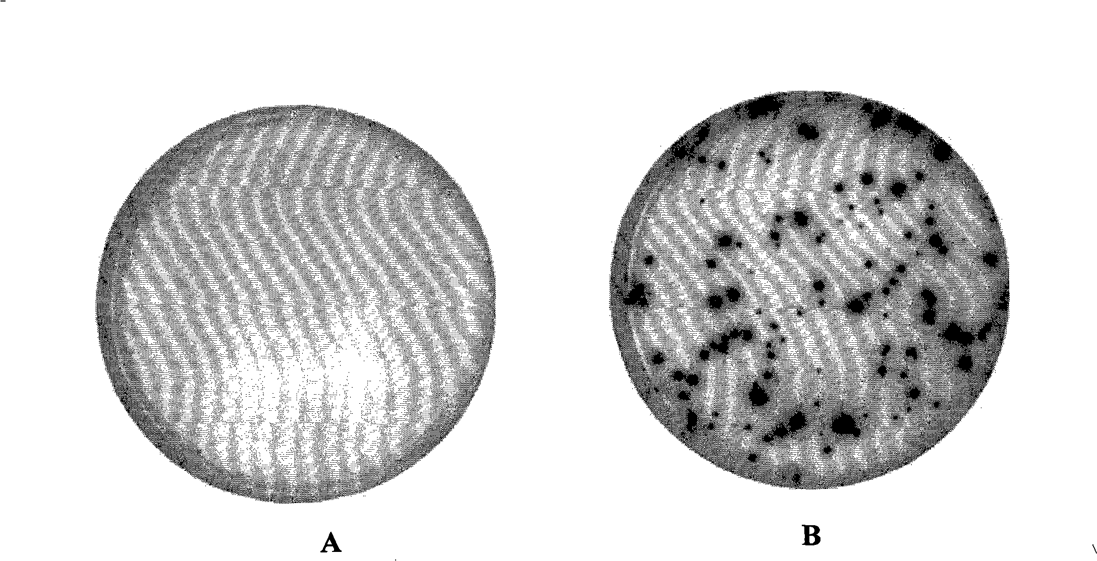
[0138] Detection of IFN-γ secretion by ELISPOT method

[0139] ● method one

[0140] 1) Pre-coat overnight with 10ug / ml IFN-γmAB on a 96-well PVDF plate (Millipore)

[0141] 2) Wash twice with medium RPMI 1640, block with RPMI 1640+10% FBS at 4°C for 1 hour

[0142] 3) Add 100ul to each well with a concentration of 5×10 5 PBMC cells / ml. In the experimental group, proteins or polypeptides were added to the wells with a final concentration of 10ug / ml; PHA with a final concentration of 5ug / ml was added to the positive control wells; 100ul AIV-M was added to the negative control wells.

[0143] 4) Incubate for 22 hours in a 37°C, 5% CO2 incubator.

[0144] 5) The cell solution was discarded, and the plate was washed 5 times with PBS containing 0.05% Tween20, 250ul / well each time.

[0145] 6) Add 50ul of freshly prepared 1:200 alkaline phosphatase-labeled anti-IFN-γ mAb, and incubate at 4°C for 1 hour.

[0146] 7) Wash the plate 5 times with PBS buffer containing 0.05% Twee...


PUM


Abstract

The invention belongs to the biomedical detection field, in particular relates to a reagent and method used for detecting activity tuberculosis and latent tuberculosis infection; based on the genomic principle, the invention discloses a novel detection reagent for mycobacterium tuberculosis, containing protein or polypeptide which is represented by SEQ ID1-2, 4-5, 8-28; the method uses one or a plurality of SEQ ID 1-28 protein or polypeptide to contact T cells of a mycobacterium tuberculosis host, and detects cytokine released from the T cells; the method can detect the tuberculosis and latent infection effectively and is not interfered by BCG vaccine at the same time; the invention also discloses a diagnostic reagent kit and other application based on the protein or polypeptide and the method; compared with the T-SPOT of the prior art, the invention can improve detectable rate obviously under the condition that the specificity is not reduced; the reagent kit has cheap price, and the cost is 1 / 5 to 1 / 10 of that of the T-SPOT reagent, thus being beneficial to being popularized in the developing countries and poor areas.

Description

technical field [0001] The invention belongs to the field of biomedical testing, and relates to a detection method of microbial molecular biology, in particular to a reagent and a method for detecting active tuberculosis and tuberculosis latent infection. Background technique [0002] The causative agent of tuberculosis is Mycobacterium tuberculosis, which belongs to the genus Mycobacterium. Tuberculosis has become a major public health problem today. According to relevant reports, there are about 8-10 million new tuberculosis cases in the world every year, and 2-3 million people lose their lives every year. Tuberculosis is the number one infectious disease that causes adult deaths. According to WHO reports, one-third of the world's population (about 2 billion) has been infected with Mycobacterium tuberculosis. If no measures are taken, about 300 million people will be infected by Mycobacterium tuberculosis in the past 10 years. According to the fourth national epidemiolog...

Claims


Application Information

Patent Timeline
IPC IPC(8): G01N33/569G01N33/68
Inventor 张文宏陈嘉臻张颖王洪海王九玲莫凌邵凌云
Owner AFFILIATED HUSN HOSPITAL OF FUDAN UNIV
Learn More