Detection method and reagent kit of anti-keratin antibody
An anti-keratin, detection method technology, applied in biological testing, material inspection products, fluorescence/phosphorescence, etc., can solve the problems of weak fluorescence intensity, missed detection, etc., achieve increased fluorescence intensity, convenient use, improved resolution and The effect of sensitivity
- Summary
- Abstract
- Description
- Claims
- Application Information
AI Technical Summary
Problems solved by technology
Method used
Image
Examples
Embodiment 1
[0029] The detection method of embodiment 1 anti-keratin antibody
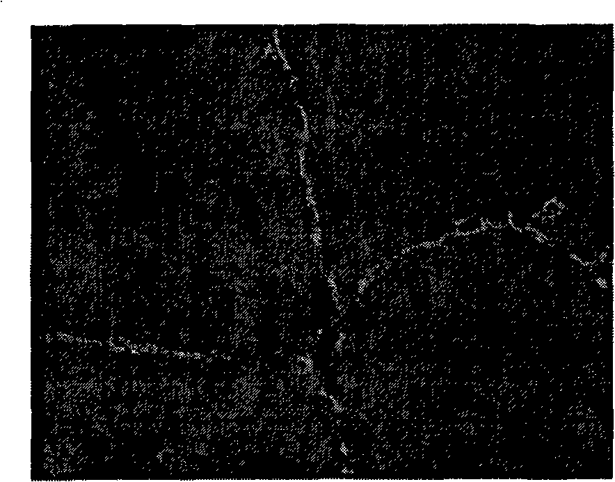
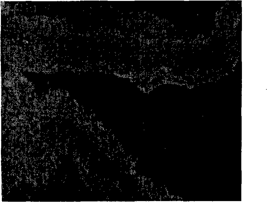
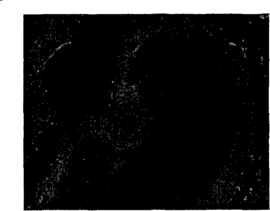
[0030] During detection, add 25 μl of 1:10 diluted serum to be tested in the reaction area of the biological sheet, incubate in a wet box at 37°C for 30 minutes, rinse with PBS, spin dry, add 25 μl of complement, incubate in a wet box at 37°C for 30 minutes, rinse with PBS, Shake dry, then add fluorescein-labeled anti-C 3 c antibody 25 μl, incubated at 37°C for 30 minutes, rinsed with PBS, dried, and mounted with buffered glycerol. Mounting is not required for direct detection. The slides were observed under a fluorescent microscope, and the typical regular linear or lamellar fluorescence in the stratum corneum was positive.
[0031] In order to better understand the present invention, the positive effect of the present invention aspect detection anti-keratin antibody is further illustrated by the result that two kinds of detection methods draw during clinical detection below:
[0032] The selected cases ...
Embodiment 2
[0042] Example 2 Kit
[0043] 1. Box body
[0044] The kit of the present invention is for one-time use. The box body 1 is in the shape of a hexahedron made of cardboard and consists of a box bottom 5 and a box cover 2 . The bottom of the box is connected with the lid as a whole. Bottom of the box 5 is provided with a bottle holder 3 at the long side where the bottom of the box 5 links to each other with the lid 2, and the bottle holder is provided with 7 holes 4 for placing bottles 10 that various reagents are housed. In the middle of the bottom of the box 5 there is a baffle 7 extending from the bottle holder 3 to the other long side, dividing the bottom of the box 5 into two parts, one of which is placed with a buffer and a cover, and the other part is placed with a carrier sheet with a packaging bag 6 13 and instructions (not shown). The length, width and height of the box body match the shape of the contained items.
[0045] 2. Items equipped with the kit
[0046] 1...
Embodiment 3
[0056] Embodiment 3 kit experimental operation steps
[0057] Preparation: Take the slides out of the kit and let it warm to room temperature.
[0058] Dilution: first dilute 50ml of concentrated phosphate buffer solution with distilled water to 1L, the phosphate content of the diluent is 10.2g, pH=7.2. Add 2ml Tween 20 into the diluted phosphate buffered saline (PBS-Tween 20), and stir well. Dilute the serum to be tested with PBS-Tween 20 at a ratio of 1:10 (positive and negative control serum do not need to be diluted), and mix well before use. Reconstitute 100 μg of powdered complement with 50 μl of distilled water into a complement application solution.
[0059] Loading: Add 25ul of diluted serum dropwise to the reaction area of the slide to avoid air bubbles. For the first incubation: place the slide with the bioflake facing up, cover the reaction area with a cover slip, and the reaction starts immediately. Make sure the specimen is in contact with the biochip and i...
PUM
 Abstract
Description
Claims
Application Information
 - R&D
- Intellectual Property
- Life Sciences
- Materials
- Tech Scout
- Unparalleled Data Quality
- Higher Quality Content
- 60% Fewer Hallucinations
Browse by: Latest US Patents, China's latest patents, Technical Efficacy Thesaurus, Application Domain, Technology Topic, Popular Technical Reports.
© 2025 PatSnap. All rights reserved.Legal|Privacy policy|Modern Slavery Act Transparency Statement|Sitemap|About US| Contact US: help@patsnap.com


