Mosaic type adenoviral vector for transfecting pig skin and use thereof
An adenovirus and chimeric technology, applied in the field of biomedicine, can solve the problems of not meeting long-term coverage of wounds, slowing down humoral immune rejection, etc., and achieve the effect of improving transplant survival time and transfection efficiency
- Summary
- Abstract
- Description
- Claims
- Application Information
AI Technical Summary
Problems solved by technology
Method used
Image
Examples
Embodiment 1
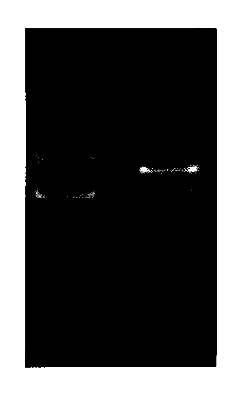
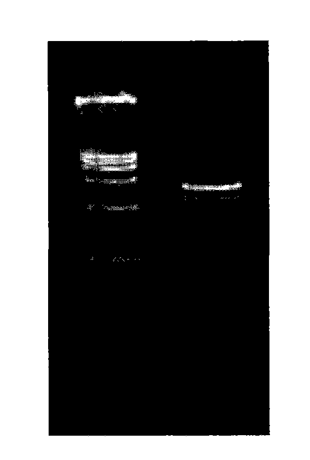
[0026] Example 1: Preparation of Ad5F35-CTLA4Ig-CD40Ig chimeric adenovirus vector
[0027] Step 1: Construction of CTLA4Ig fusion protein expression vector
[0028] The pUC19-CTLA4Ig plasmid was constructed according to Example 1 disclosed in the patent application No. 00103528 and the title of the invention is "a kind of allogeneic skin transfected with recombinant gene". The CTLA4Ig recombinant gene expression series is shown in SEQ ID NO:1.
[0029] Step 2: Construction of CD40Ig fusion protein expression vector
[0030] 1), routine method is isolated mononuclear cell from the peripheral blood of people;
[0031] 2), conventional methods extract the RNA of mononuclear cells; and reverse transcribe into cDNA;
[0032] 3), the extracellular region of CD40 is amplified by PCR with specific primers; and the product is cloned into T vector;
[0033] The sequence of primer 1 is shown in SEQ ID NO:5, and the sequence of primer 2 is shown in SEQ ID NO:6.
[0034] Primer 1: 5'-...
Embodiment 2
[0063] Example 2: Real-time quantitative PCR detection of target gene transfected with porcine skin tissue by Ad5F35-CTLA4Ig-CD40Ig chimeric adenovirus vector in vitro
[0064] 1. Preparation of pigskin slices: In the ultra-clean workbench, cut the pigskin into small pieces of 2cm×2cm, disinfect with 5% povidone iodine for 10 minutes, wash with a large amount of sterilized normal saline, and set aside.
[0065] 2 Transfection: adjust the Ad5F35-CTLA4Ig-CD40Ig chimeric adenovirus vector titer to 10 with DMEM medium 8 PFU, Ad5-CTLA4Ig adenoviral vector titer to 5×10 8 PFU, take the same area of pig skin slices (6-month-old Bama pigs), add the same volume of 10 8 PFU Ad5F35-CTLA4Ig chimeric adenovirus solution and 5×10 8 PFU Ad5-CTLA4Ig adenovirus solution was placed in a cell culture incubator at 37°C for culture. Another pig skin with the same area and the same source was transfected and cultured in a virus-free medium as a control.
[0066] 3. Real-time quantitative PCR ...
Embodiment 3
[0085] Example 3. Detection of Ad5F35-CTLA4Ig-CD40Ig chimeric adenovirus vector-mediated immunosuppressive function in vitro
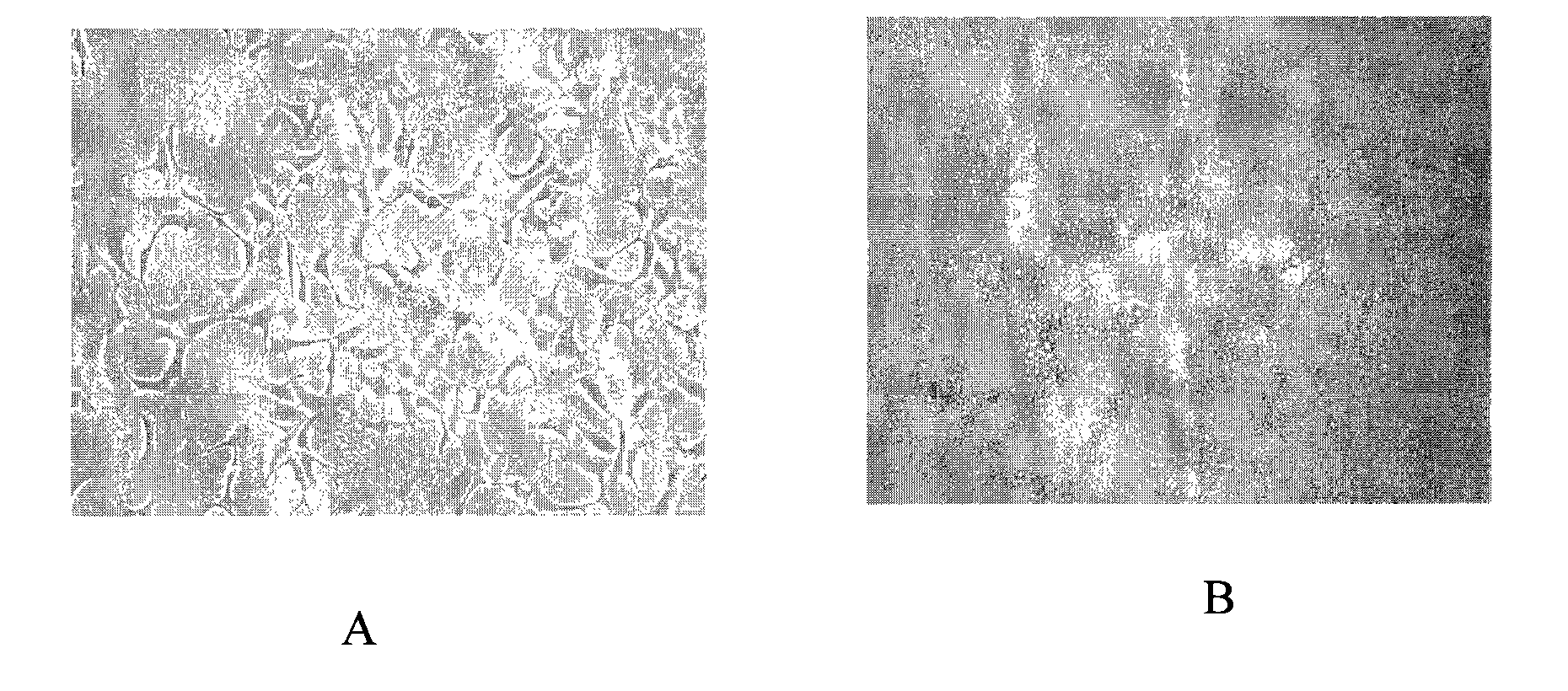
[0086] Pig fibroblasts were 1.0×10 4 cell / well (96-well plate) was subcultured in 100 μl high-glucose DMEM+10% fetal bovine serum medium, and the culture conditions were 37°C, 5% CO 2 , 4-12 hours after the cells adhered to the wall, wash 3 times with 0.01M / LPBS, Ad5F35-CTLA4Ig-CD40Ig chimeric adenovirus vector, Ad5-CTLA4Ig adenovirus vector were transfected at MOI=100, 100μl for 2 hours, blotted transfection solution. Lymphocytes were separated using human lymphocyte separation medium. Resuspend 2.0×10 in 200 μl RPMI 1640 complete medium containing 10% calf serum 5 cells / well (96-well plate) were co-cultured with fibroblasts. After 48h, centrifuge at 1000rpm×5min, add 20μl of 0.5mg / ml MTT solution, continue to incubate for 4h, add 100μlDMSO after suction and incubate on an air-bath shaker at room temperature for 10min, measure the OD value at 570n...
PUM
 Abstract
Description
Claims
Application Information
 - R&D
- Intellectual Property
- Life Sciences
- Materials
- Tech Scout
- Unparalleled Data Quality
- Higher Quality Content
- 60% Fewer Hallucinations
Browse by: Latest US Patents, China's latest patents, Technical Efficacy Thesaurus, Application Domain, Technology Topic, Popular Technical Reports.
© 2025 PatSnap. All rights reserved.Legal|Privacy policy|Modern Slavery Act Transparency Statement|Sitemap|About US| Contact US: help@patsnap.com


