Collagen-based microspheres and methods of preparation and use thereof

A technology of microspheres and collagen, applied in the direction of microsphere preparation, microcapsule preparation, capsule delivery, etc., can solve the problems affecting the interaction between drug and matrix, affecting the initial burst release and release rate, etc.

Active Publication Date: 2010-10-20
THE UNIVERSITY OF HONG KONG
View PDF3 Cites 7 Cited by
  • Summary
  • Abstract
  • Description
  • Claims
  • Application Information

AI Technical Summary

Problems solved by technology

Drug factors such as surface charge, hydrophobicity, loading and solubility in the continuous phase of the drug may affect the drug-matrix interaction and thus the initial burst and release rate
Matrix f

Method used


Image

  • Collagen-based microspheres and methods of preparation and use thereof
  • Collagen-based microspheres and methods of preparation and use thereof
  • Collagen-based microspheres and methods of preparation and use thereof

Examples


Embodiment 1

[0090] Example 1. Preparation of Collagen Microspheres Incorporated into BSA or Myoglobin

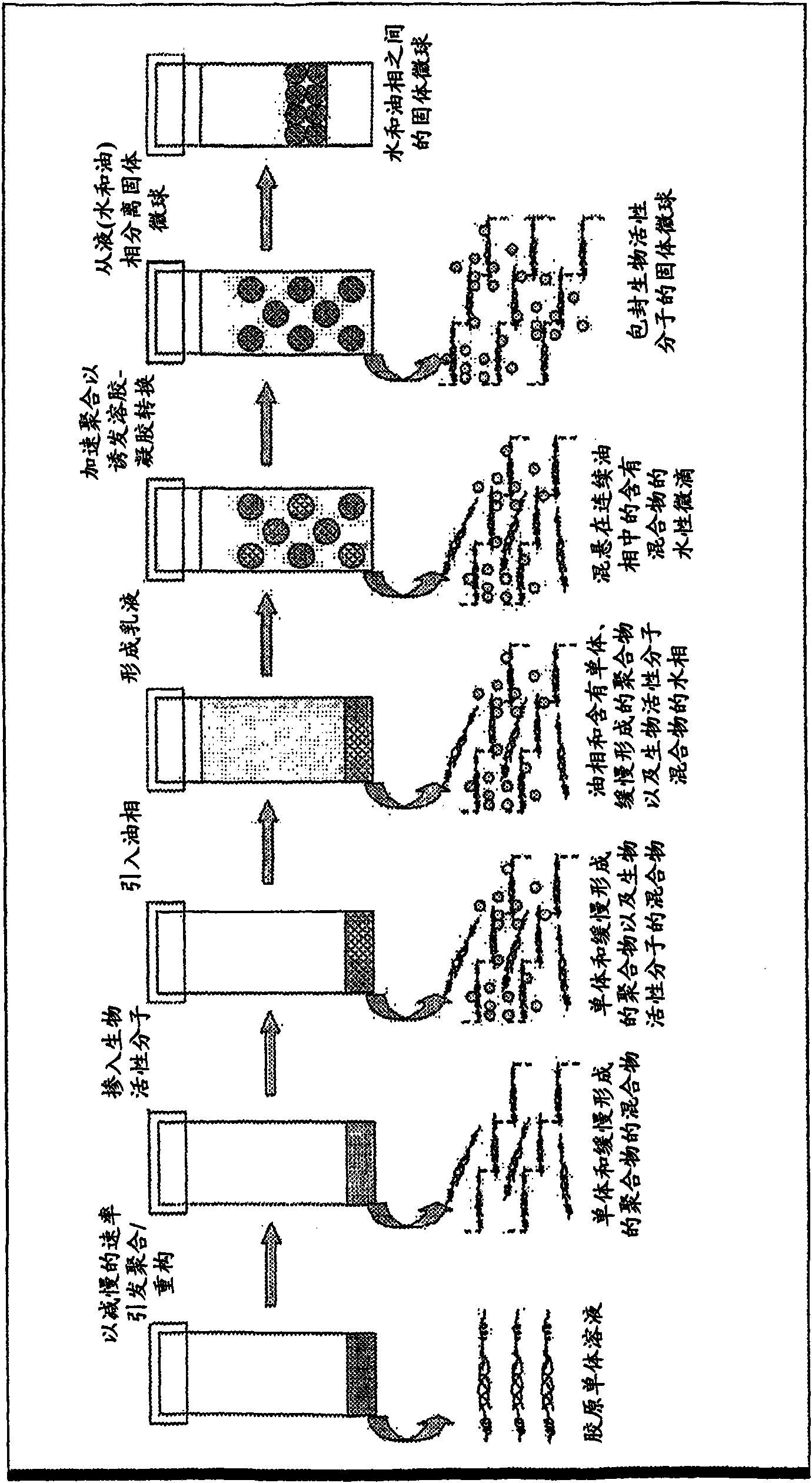
[0091] A 0.02N acetic acid solution with a concentration of 7mg / ml rat tail collagen type I was neutralized with 1N sodium hydroxide in the presence of 10X phosphate buffer to make the final concentration 0.5X. The gelled mixture was placed in an ice-water bath at 4°C to slow the rate of polymerization for 1 minute. Sample proteins such as BSA and myoglobin can be incorporated. BSA (4 mg) was added to the mixture and mixed well.

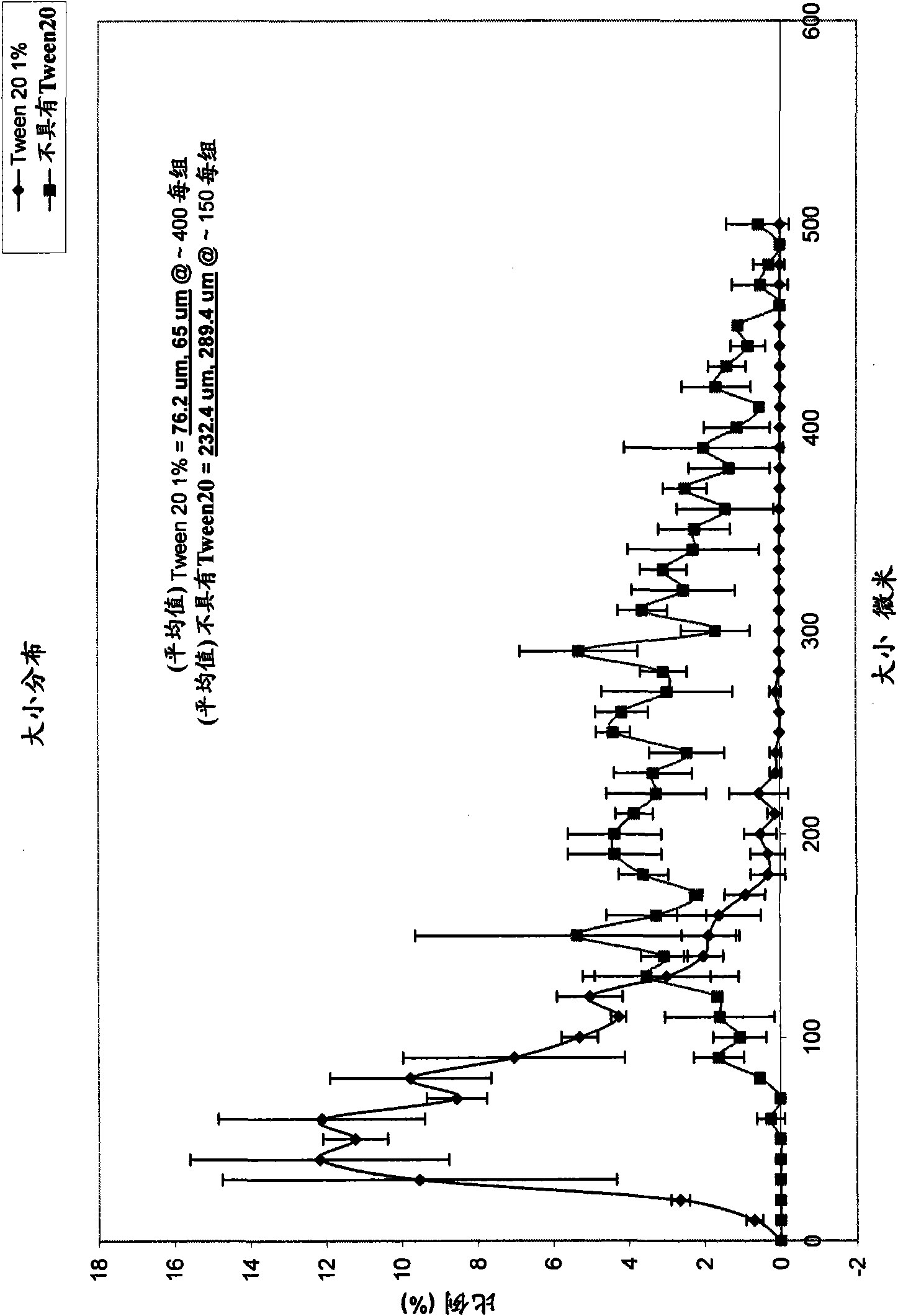
[0092] A 1:1 mixture of olive oil and silicone oil was added to the aqueous gelling mixture in a volume ratio of 6:1. The nonionic surfactant Tween Tween 20 was added to the aqueous phase before emulsification. Place the container of the mixture on the mixing device. The mixture was agitated for 30 seconds at maximum speed (3000 rpm). The formed emulsion was then placed in a 37°C water bath to accelerate polymerization. The mixture was incubated for 30 m...

Embodiment 2

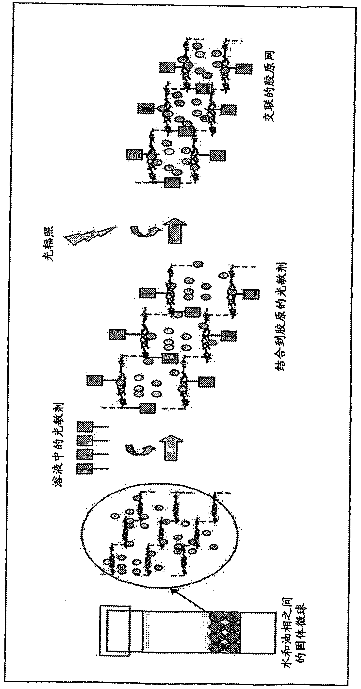
[0093] Example 2. Photochemical crosslinking of collagen microspheres

[0094] Microspheres obtained from the procedure described in Example 1 were soaked in a solution of rose bengal (photocrosslinking reagent, "PC") at a concentration of 0.001% (w / v) for 10 minutes. Discard excess rose bengal and rinse beads. The microspheres were resuspended in water and placed in 4-well culture dishes. Using 514nm argon laser to 0.02W / cm 2 Irradiate the microspheres for 100 sec. The microspheres were rinsed in water immediately after irradiation. The microspheres are then ready for injection or subsequent experiments.

Embodiment 3

[0095] Example 3. Dehydration of Collagen Microspheres

[0096]Soak the microspheres in 100% ethanol for 30 minutes and repeat three times to extract water from the microspheres. Gradually increasing ethanol concentration was used to improve the surface smoothness of the microspheres. Collagen microspheres were soaked in 50% v / v ethanol for 20 minutes, twice in 70% ethanol for 30 minutes, twice in 80% ethanol, twice in 90% ethanol and twice in 100% ethanol.


PUM


Abstract

A method of manufacture of ECM microparticles incorporating bioactive molecules for drug delivery has been developed, using a modified emulsification method or a water-in-oil-phase-separation method. The microspheres are photochemically crosslinked to control the release of the bioactive molecules for better drug delivery usage without compromising the biocompatibility of the crosslinked structures. The method uses mild fabrication conditions and simple processes, no toxic chemical crosslinking reagent, which may cause cytotoxicity and calcification after implantation, no organic solvents, which may reduce drug availability and bioactivity, and no vigorous stirring action, which may fragmentize material with poor shape and mechanical stability and thus destabilize the emulsion. The resulting microparticles or microspheres are of controlled size, controlled release, highly biocompatible, and useful for drug delivery as well as cell culture.

Description

field of invention [0001] The present application generally relates to microparticles formed from extracellular matrix materials that provide controlled release and desired mechanical properties, and methods of preparation and use thereof. [0002] Cross References to Related Applications [0003] This application claims the benefit and priority of U.S.S.N. 60 / 948,336, filed July 6, 2007, and is a continuation of U.S.S.N. 11 / 225,108, filed September 14, 2005, which claims September 14, 2004 Benefit and priority of U.S.S.N. 60 / 609,600 filed. The disclosures in the applications listed above are incorporated herein by reference. Background of the invention [0004] Microsphere-based drug delivery systems are advantageous due to their injectability and versatility in controlling the release mode of the loaded drug (Sinha and Trehan, J. Control. Release, 90(3): 261-80 (2003) ). This reduces the invasiveness of multiple injections. Biocompatibility and biodegradability are es...

Claims


Application Information

Patent Timeline
IPC IPC(8): B01J13/14A61K9/52A61K9/50
CPCA61L27/3633A61K9/1694B01J13/14A61K9/1658B01J13/06
Inventor 陈佩陈卓铭苏国辉
Owner THE UNIVERSITY OF HONG KONG
Why Eureka
Try Eureka