Immunochromatographic test strip for rapidly detecting acute pancreatitis and preparation method thereof

A technology of acute pancreatitis and immunochromatography, applied in the field of medical testing, can solve the problems of high cost, complicated preparation process of colloidal gold labeling, sensitivity to be improved, etc., to achieve simple operation, easy-to-read results, improved sensitivity and simplicity Effect

Active Publication Date: 2011-06-08
GUANGZHOU WONDFO BIOTECH
View PDF3 Cites 6 Cited by
  • Summary
  • Abstract
  • Description
  • Claims
  • Application Information

AI Technical Summary

Problems solved by technology

The preparation process of colloidal gold labeling is complicated, and the cost is high, and the sensitivity needs to be improved

Method used


Image

  • Immunochromatographic test strip for rapidly detecting acute pancreatitis and preparation method thereof
  • Immunochromatographic test strip for rapidly detecting acute pancreatitis and preparation method thereof

Examples


Embodiment 1

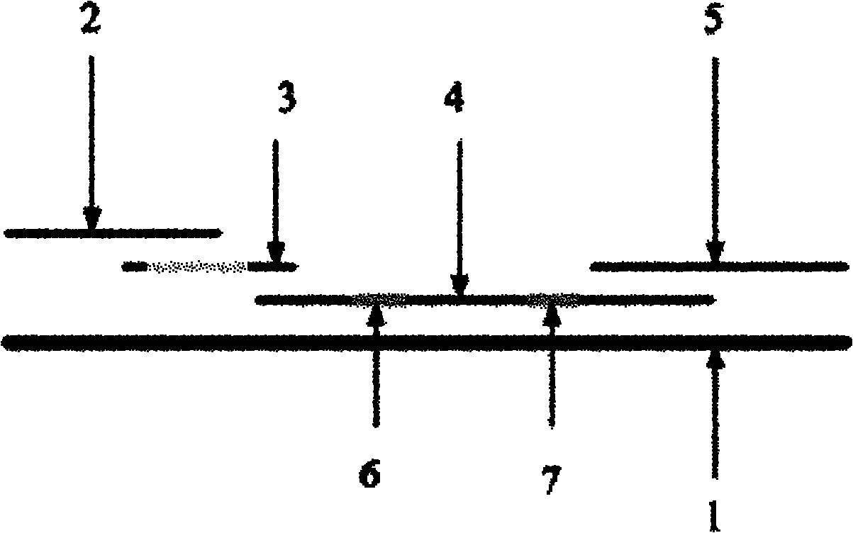
[0037] Such as figure 1 As shown, it is a structural schematic diagram of a quick detection test strip of acute pancreatitis immunochromatography according to the present invention. It is formed by lapping and pasting on the substrate 1 in sequence, and the glass fiber membrane marking pad 3 is coated with a biotin-avidin-color latex complex-labeled trypsinogen-2 high-specificity monoclonal antibody, nitric acid The cellulose-coated membrane 4 includes a detection zone 6 and a control zone 7, the detection zone 6 is coated with another trypsinogen-2 specific monoclonal antibody with a different epitope from the monoclonal antibody labeled on the glass fiber membrane marker pad 3 , the control region 7 was coated with anti-mouse antibody.

[0038] In this embodiment, the ratio of biotin:avidin:color latex is 1:1:1~2:2:3, and trypsinogen-2 labeled with biotin-avidin-color latex complex is highly specific The amount of the monoclonal antibody sprayed on the glass fiber membrane...

Embodiment 2

[0053] The detection test strip structure in this embodiment is all identical with embodiment 1.

[0054] In this embodiment, the ratio of biotin:avidin:color latex is 1:1:1~2:2:3, and trypsinogen-2 labeled with biotin-avidin-color latex complex is highly specific The amount of the monoclonal antibody sprayed on the glass fiber membrane marker pad was 35 μl / cm. The concentration of trypsinogen-2 specific monoclonal antibody on the nitrocellulose-coated membrane is 7 μg / ml, and the consumption in the detection zone is 20 μl / 35cm; the concentration of the anti-mouse antibody is 7 μg / ml, in the The amount used in the control area was 20 μl / 35cm.

[0055] In the preparation method of this example, except that the weight ratio of avidin to colored latex in step B is 2:1, and the volume ratio of biotin-labeled antibody to avidin-colored latex complex in step D is 7:1, the rest All are identical with embodiment 1, and using method is also identical with embodiment 1.

Embodiment 3

[0057] The detection test strip structure in this embodiment is all identical with embodiment 1.

[0058] In this embodiment, the ratio of biotin:avidin:color latex is 1:1:1~2:2:3, and trypsinogen-2 labeled with biotin-avidin-color latex complex is highly specific The amount of the monoclonal antibody sprayed on the glass fiber membrane marker pad was 48 μl / cm. The concentration of trypsinogen-2 specific monoclonal antibody on the nitrocellulose-coated membrane is 13 μg / ml, and the consumption in the detection area is 20 μl / 35cm; the concentration of the anti-mouse antibody is 13 μg / ml, and the The amount used in the control area was 20 μl / 35cm.

[0059] In the preparation method of this example, except that the weight ratio of avidin to colored latex in step B is 3:1, and the volume ratio of biotin-labeled antibody to avidin-colored latex complex in step D is 10:1, the rest All are identical with embodiment 1, and using method is also identical with embodiment 1.

[0060] ...


PUM

PropertyMeasurementUnit
diameteraaaaaaaaaa

Abstract

The invention discloses an immunochromatographic test strip for rapidly detecting acute pancreatitis and a preparation method thereof. The test strip is formed by sticking a sample pad, a glass fiber membrane labeling pad, a nitrocellulose coating membrane and absorbent paper to a substrate in sequence through lap joint, wherein a highly specific monoclonal antibody of trypsinogen-2 labeled by a biotin-avidin-colored latex complex is coated on the glass fiber membrane labeling pad; the nitrocellulose coating membrane comprises a detection region and a control region; the detection region is coated by another specific monoclonal antibody of trypsinogen-2 with epitope different from that of the labeled monoclonal antibody on the glass fiber membrane labeling pad; and the control region is coated by an anti-mouse antibody. Compared with radioimmunoassay and enzyme linked immunosorbent assay (ELISA), the test strip has the advantages of safety, simpleness and convenience in operation, suitability for single human / sample detection, rapidness and the like, dispenses with special skills, is free of trauma, is easy in result reading and can realize timely in-situ and family self-detectionof acute pancreatitis.

Description

technical field [0001] The invention belongs to the field of medical examination, in particular, the invention relates to a rapid detection test strip of acute pancreatitis immunochromatography and a preparation method thereof. Background technique [0002] Acute pancreatitis, especially severe acute pancreatitis, is a serious disease that threatens people's health. It has been listed as one of the three acute abdomen diseases. Most acute pancreatitis is mild, and about 20% of patients are severely necrotic. The mortality rate of acute severe pancreatitis can reach 40%, so the early diagnosis of acute pancreatitis is of great significance to the treatment and prognosis of acute severe pancreatitis. [0003] CT is one of the most accurate methods for diagnosing acute pancreatitis, but it is not cost-effective due to its high cost. Traditional blood and urine amylase are widely used as indicators for clinical diagnosis and monitoring of the course of acute pancreatitis. Howev...

Claims


Application Information

Patent Timeline
Patent Type & Authority Applications(China)
IPC IPC(8): G01N33/577G01N33/558G01N33/532
Inventor 王继华李凯
Owner GUANGZHOU WONDFO BIOTECH
Why Eureka
Try Eureka