Dual-mode optical imaging probe and preparation method thereof

An optical imaging, dual-mode technology, applied in the fields of nanomaterials and bioanalytical chemistry, can solve the problems of further improvement of sensitivity, stability and biocompatibility, complicated preparation methods, etc., and achieve surface-enhanced Raman signals and fluorescence signals. , Good repeatability and high sensitivity

Inactive Publication Date: 2011-09-07
SOUTHEAST UNIV
View PDF1 Cites 22 Cited by
  • Summary
  • Abstract
  • Description
  • Claims
  • Application Information

AI Technical Summary

Problems solved by technology

Although there have been a lot of reports on the structure and preparation methods of SERS probes, there are not many probes suitable for living

Method used


Image

  • Dual-mode optical imaging probe and preparation method thereof
  • Dual-mode optical imaging probe and preparation method thereof
  • Dual-mode optical imaging probe and preparation method thereof

Examples


Embodiment 1

[0023] Examples 1 and 2 use gold nanoparticles as the SERS substrate, 5,5-dithiobis(2-nitrobenzoic acid) DTNB as the Raman marker, and Rhodamine 6G as the fluorescent material for illustration. .

[0024] Example 1

[0025] To prepare a dual-mode optical imaging label using rhodamine 6G as a fluorescent material and DTNB molecules as a Raman marker, the method includes the following steps:

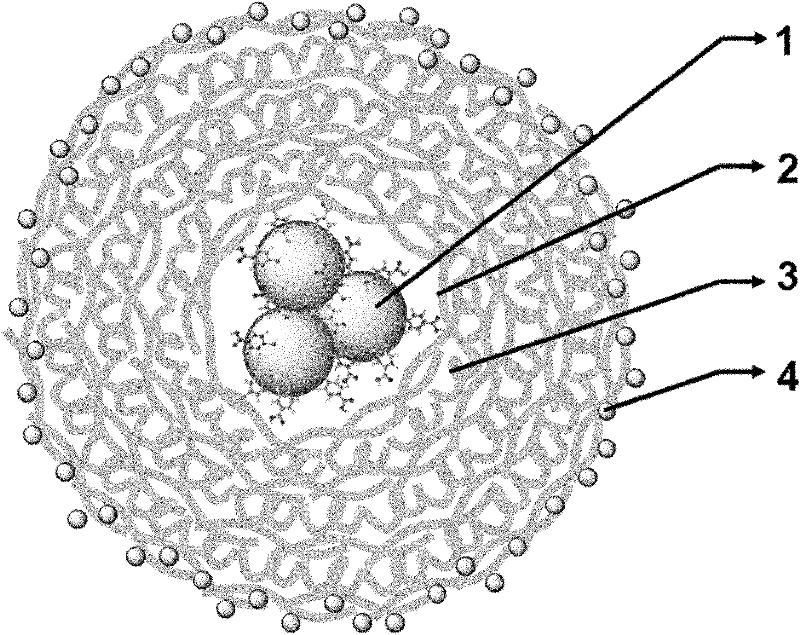
[0026] 1) The experiment uses the method reported by Frens to prepare gold colloidal solution. Under vigorous stirring, 4 mL of citric acid aqueous solution (concentration of 1%) was added into boiling 100 mL of chloroauric acid aqueous solution (concentration of 10 -4 g / mL), continuously stirred and kept boiling for 20 minutes to obtain an aqueous solution of gold nanoparticles, and the average particle diameter of the particles was 15nm. Add 0.2 mL of the aqueous solution of the Raman marker DTNB into 20 mL of the gold nanoparticle solution to a final concentration of 10 -5 ~10 -6 M...

Embodiment 2

[0034] Fluorescence and SERS properties of dual-mode optical imaging probes in living cells (a probe with rhodamine 6G as the fluorescent material and DTNB molecule as the Raman marker)

[0035] 1) Place cervical cancer cells (HeLa) in culture medium for in vitro culture (37°C, 5% CO 2 ). After 24 hours, the aqueous solution of the dual-mode imaging probe was added to the cell culture medium in a volume ratio (3:1), shaken gently, and placed in the incubator again. The probe enters the interior of the cell by being phagocytized by the cell. After 1.5 hours, the medium was aspirated, and the cells were washed 3 times with phosphate buffered saline (PBS) to remove the dual-mode imaging probes that were not phagocytized by the cells and remained in the medium, ready for use.

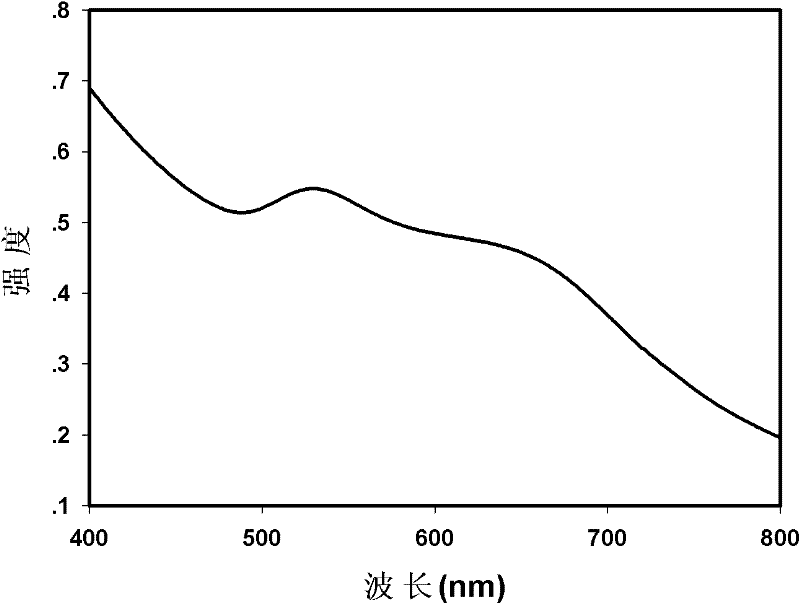
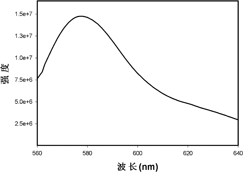
[0036] 2) Place the cells washed with buffer solution on the stage of a confocal microscope, use 543nm as the excitation wavelength and 560nm-640nm as the receiving wavelength range to obtain the cell flu...

Embodiment 3

[0040] Using silver nano-aggregates as a SERS substrate to prepare a dual-mode optical imaging label using cadmium telluride (CdTe) quantum dots as a fluorescent material and 4-mercaptobenzoic acid molecules (4MBA) as a Raman marker, the method includes the following steps :

[0041] 1) The experiment adopts the method reported by Lee and Meisel to prepare silver colloid solution. will be 1.0×10 -2 M silver nitrate solution was added to deionized water according to the volume ratio of 1:10 to the silver colloid solution to be obtained, stirred and heated to boiling. Add 1% sodium citrate solution into the boiling silver nitrate solution according to the volume ratio of 1:50 to the silver colloid solution to be obtained, keep stirring and heat and boil for 40 minutes to obtain the silver colloid solution. Add 0.2 mL of 4MBA in water to 20 mL of the silver nanoparticles solution to a final concentration of 10 -5 ~10 -6 M, mixing and stirring for 30 minutes, it can be observe...


PUM


Abstract

The invention discloses a dual-mode optical imaging probe and a preparation method thereof. The dual-mode optical imaging probe comprises nano particles which are dispersed in a water solution, each nano particle comprises a core body and a wrapping layer, wherein the core body is a gold or silver nano aggregate; the wrapping layer is multilayer PDDA (Poly (Diallyldimethylammonium Chloride)) and PSS (Poly Sodium Styrenesulfonate) which are alternately adsorbed; and the outermost layer of the wrapping layer is PDDA with fluorescent materials absorbed. The preparation method of the dual-mode optical imaging probe comprises the following steps of: preparing the Raman marker-induced gold or silver nano aggregate; then adding a PDDA water solution to a water solution of the Raman marker induced gold or silver nano aggregate, stirring, and then alternately adsorbing PSS and PDDA for multiple times; then adding the fluorescent materials and the PDDA water solution, and the like. The dual-mode optical imaging probe has both surface enhanced Raman signals and fluorescent signals, high sensitivity, easiness of optical probe multifunction realization and important application values in the fields of drug targeted transportation, biological sensing, detection, and the like; and the preparation method of the dual-mode optical imaging probe has the advantages of easiness of operation, good repeatability, low cost and environmental friendliness.

Description

technical field [0001] The invention relates to the fields of nanomaterials and bioanalytical chemistry, in particular to a dual-mode optical imaging probe and a preparation method thereof. The dual-mode optical imaging probe integrates fluorescence and surface-enhanced Raman scattering (SERS) signals; The preparation method has the advantages of simple operation, good repeatability, low cost and environmental friendliness. Background technique [0002] Optical imaging technology can visually display gene expression and cell activity in living organisms. It is a powerful means in molecular and cell imaging technology and is being more and more widely used in the fields of medical and biological research. Compared with other imaging techniques in living animals, such as ultrasound (ultrasound), computed tomography (CT), magnetic resonance imaging (magnetic resonance imaging, MRI), positron-emissiontomography (PET) , Optical imaging has many unique advantages, such as easy op...

Claims


Application Information

Patent Timeline
IPC IPC(8): G01N21/64G01N21/65
Inventor 王著元崔一平宗慎飞陈辉
Owner SOUTHEAST UNIV
Why Eureka
Try Eureka