

Monoclonal antibody of anti-human PIGF (placental growth factor) protein as well as preparation method and application thereof
A monoclonal antibody and protein technology, applied in the direction of anti-animal/human immunoglobulin, animal/human protein, botanical equipment and methods, etc., can solve the problem of inability to achieve neutralization and inhibition of angiogenesis, inability to recognize or cover VEGF other antigenic sites and other issues
- Summary
- Abstract
- Description
- Claims
- Application Information
AI Technical Summary
Problems solved by technology
Method used
Image

Examples
Embodiment 1
[0046] Example 1: Preparation of immune antigen: expression preparation and in vitro activity identification of recombinant human PlGF131 protein
[0047] The expression and preparation of recombinant human PlGF131 protein is prepared by the method described in the Chinese invention patent application with the application number 200810018884.8 and the title of the invention "Expression production, separation and purification of recombinant placental growth factor and its chemical labeling".
[0048] The gene fragments encoding the complete open-reading frame (ORF) of human PlGF131 were amplified from the cDNA library of human lung tissue cells by PCR technology, which were identified correctly by sequence determination and treated with restriction endonucleases. , and clone it into the yeast expression vector pPic9K (Invitrogen Company) vector. The expression plasmid pPic9K-PlGF131 was obtained. After electrotransformation of Pichia pastoris, high-efficiency expression engine...
Embodiment 2
[0051] Example 2: Establishment and screening identification of a hybridoma cell line stably secreting a monoclonal antibody against human PlGF protein
[0052] Step 1. Animal immunization:
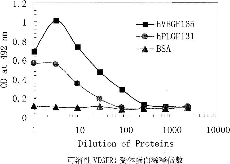
[0053] The recombinant human PlGF131 protein purified in Example 1 above was mixed with complete Freund's adjuvant (purchased from Sigma, USA), and injected subcutaneously into C57BL / 6 mice (Shanghai Slack Experimental Animal Co., Ltd., 100ul / only, A total of 10ug PlGF131 protein). 2-3 weeks after the first immunization, the mice were boosted by multi-point subcutaneous injection of a mixture of human PlGF131 protein and incomplete adjuvant (purchased from Sigma, USA). After 2-3 times of booster immunization, take a small amount of mouse serum, and use a 96-well microtiter plate coated with human PlGF131 protein to detect the titer of anti-PlGF131 protein antibody in mouse serum by ELISA method. Splenocytes were used for the next step of cell fusion.
[0054] Step 2, cell fusion:
[0...
Embodiment 3
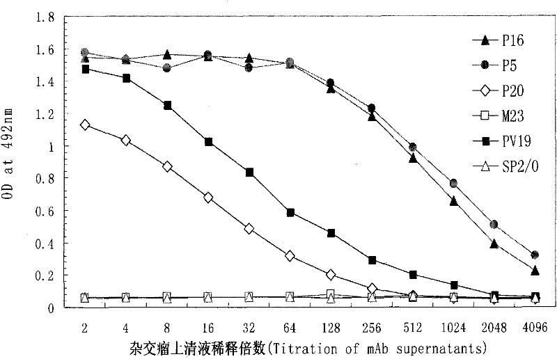
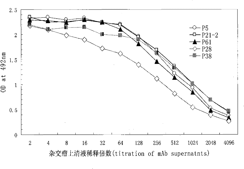
[0063] Example 3. In vitro separation, purification and detection of monoclonal antibodies against human PlGF protein
[0064] Step 1: amplify and culture the hybridoma cells established in Example 2 above in a serum-free medium until the cell concentration reaches 10 5 Stop adding liquid when the cell culture medium is above / ml, continue culturing until the cell culture medium turns yellow, collect the culture medium, centrifuge at 1500rpm for 10 minutes, filter the supernatant through a 0.45μm filter membrane, and store it at 4°C or -20°C for later use or directly use The next step is the separation and purification of monoclonal antibodies.
[0065] Step 2: Separation and purification of monoclonal antibody against human PlGF protein using protein-A affinity chromatography. The purification step is as follows: the supernatant of the hybridoma cell filtrate containing the monoclonal antibody against human PlGF protein is loaded on 4% agarose microparticle beads (protein-A ...
PUM
 Abstract
Description
Claims
Application Information