Preparation method of tumor detection nanoprobe

A nanoprobe and tumor technology, applied in the field of tumor detection, can solve the problems of limited resolution and difficult targeting of magnetic iron oxide nanoparticles to tumor cells or tissues

Inactive Publication Date: 2011-11-09
WUHAN INST OF VIROLOGY CHINESE ACADEMY OF SCI
View PDF1 Cites 10 Cited by
  • Summary
  • Abstract
  • Description
  • Claims
  • Application Information

AI Technical Summary

Problems solved by technology

[0006] Also, while MRI is a major imaging modality in oncology, it has limited resolution when used for molecular and cellular imaging
Moreover, MRI contrast agents alone such as magnetic iron oxide nanoparticles are difficult to target tumor cells or tissues

Method used


Image

  • Preparation method of tumor detection nanoprobe
  • Preparation method of tumor detection nanoprobe
  • Preparation method of tumor detection nanoprobe

Examples


Embodiment 1

[0048] A preparation method for detecting tumor nano probes, the steps are:

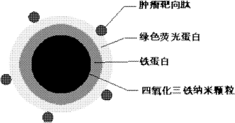
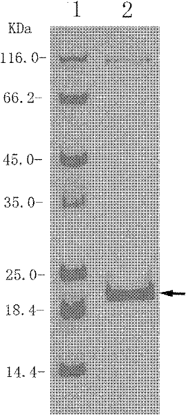
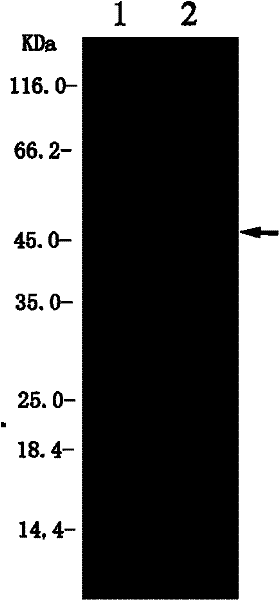
[0049] (1) Amplify and synthesize the human ferritin heavy chain gene rHF by polymerase chain reaction (PCR), insert it into the expression vector pET-28a (purchased from promega), and construct an expression plasmid pET- expressing the ferritin heavy chain. rHF, transformed into Escherichia coli BL21 (λDE3) (purchased from Promega), culture at 37°C with constant temperature, 200r / min shaking culture until OD600 is between 0.4 and 0.6, add IPTG to the culture to a final concentration of 1mmol / L After the cultures were induced and cultured at 25°C for 8 hours, the cells were collected by centrifugation at 6000g at 4°C. The cell pellet was washed once with Tris-HCl buffer (20mM Tris-HCl, 50mM NaCl, pH 8.0), and then resuspended in In 30mL Tris-HCl buffer solution, after sonication, centrifuge at 12000r / min for 30min, collect the supernatant in a 60℃ water bath for 10min, then centrifuge at 12000r / min for ...

Embodiment 2

[0054] The three-function tumor detection probe is used for specific fluorescence imaging of tumor cells. The steps are:

[0055] (1) The human glioblastoma cell line U87MG (purchased from the Chinese Type Culture Collection of Wuhan University) and the human non-small cell lung cancer cell line A549 cell line were purchased from the Chinese Type Culture Collection of Wuhan University). The surface has the specific up-regulated expression of the tumor marker αvβ3 integrin. The cells were subcultured in DMEM medium containing 10% fetal bovine serum at 37°C and 5% carbon dioxide. Passage ratio 1:2.

[0056] (2) U87MG and A549 cells are spread in a petri dish with a cover glass attached to the center, at 37 ℃, 5% carbon dioxide environment for 24-36 hours, after reaching 50% cell confluence, wash 3 times with PBS buffer. Change to binding buffer (20mM Tris-HCl, 150mM NaCl, 1mM Ca 2+ , 1mM Mg 2+ , 1% BSA, pH 7.4), add the prepared trifunctional ferritin nanoprobe to the cells to a fi...

Embodiment 3

[0059] The trifunctional tumor detection probe is used as a contrast agent for magnetic resonance imaging of tumor cells. The steps are:

[0060] (1) The human glioblastoma cell line U87MG (purchased from the Chinese Type Culture Collection of Wuhan University) and the human non-small cell lung cancer cell line A549 cell line were purchased from the Chinese Type Culture Collection of Wuhan University). The surface has the specific up-regulated expression of the tumor marker αvβ3 integrin. The cells were subcultured in DMEM medium containing 10% fetal bovine serum at 37°C and 5% carbon dioxide. Passage ratio 1:2.

[0061] (2)10 6 U87MG and A549 cells (purchased from the Chinese Type Culture Collection of Wuhan University) were washed 3 times with PBS buffer and replaced with binding buffer (20mM Tris-HCl, 150mM NaCl, 1mMCa 2+ , 1mM Mg 2+ , 1% BSA, pH 7.4), put the prepared trifunctional ferritin nanoprobe or not fused with tumor targeting peptide, but fused with fluorescent protei...


PUM

PropertyMeasurementUnit
diameteraaaaaaaaaa

Abstract

The invention discloses a preparation method of a tumor detection nanoprobe. The method comprises the following steps of: (1) constructing expression plasmids for expressing a ferritin heavy chain, expressing with colon bacillus, purifying ferritins and verifying and representing a cage structure with an electron microscope; (2) fusing a green fluorescent protein at an amino terminal of each ferritin and performing expression and purification to obtain a ferritin cage structure of which the surface expresses the fluorescent protein; (3) fusing a segment of tumor targeting small peptides RGD (Arginine-Glycine-Aspartate) at the amino terminal of a ferritin H chain of which the amino terminal is fused with the green fluorescent protein to prepare a ferritin cage structure of which the surface expresses the fluorescent protein and the tumor targeting peptides; and (4) synthesizing Fe3O5 nanoparticles in the cavity of the ferritin cage structure of which the surface expresses the fluorescent protein and the tumor targeting peptides simultaneously to obtain a multifunctional ferritin probe with tumor targeting, fluorescence and magnetism. In the nanoprobe, a tumor targeting function, a fluorescent imaging function and a magnetic resonance imaging function are fused integrally, so that synchronous detection in multiple modes can be realized.

Description

Technical field [0001] The present invention relates to the technical field of tumor detection, and more specifically to a method for preparing nanoprobes for detecting tumors, by displaying fluorescent proteins and RGD tumor-targeting short peptides on the surface of a ferritin cage structure, and synthesizing Fe in its lumen 3 O 4 Nanoparticles, prepared nanoparticle probes with three functions of tumor targeting, magnetism and fluorescence. This three-functional nanoprobe can be used for the detection of tumor cells or tumor-targeted magnetic resonance imaging (MRI) and fluorescence imaging. Background technique [0002] Cancer is one of the leading causes of human death. Although tumor biology and cancer medicine have made great progress, such as the discovery of tumor biomarkers, simple surgical procedures, and the development of radiotherapy and chemotherapy, the overall cancer survival rate has not been significant in the past two decades. improve. In order to improve th...

Claims


Application Information

Patent Timeline
Patent Type & Authority Applications(China)
IPC IPC(8): C12Q1/04A61K49/00A61K49/14
Inventor 张先恩崔宗强李可张治平危宏平
Owner WUHAN INST OF VIROLOGY CHINESE ACADEMY OF SCI
Why Eureka
Try Eureka