Activated toxic antibody kit for detecting porcine reproductive and respiratory syndrome virus

A technology for respiratory syndrome and pig breeding, which is applied in the fields of new animal disease diagnostic reagents, live virus antibody kits, and liquid-phase blocking ELISA preparation to achieve low production costs, strong specificity, and improved sensitivity and specificity Effect

Inactive Publication Date: 2012-01-04
JILIN UNIV
View PDF4 Cites 12 Cited by
  • Summary
  • Abstract
  • Description
  • Claims
  • Application Information

AI Technical Summary

Problems solved by technology

Most of the above-mentioned technicians for detecting PRRSV antibodies are aimed at structural proteins, but there are no reports on the detection methods of PRRSV non-structural protein antibodies

Method used


Image

  • Activated toxic antibody kit for detecting porcine reproductive and respiratory syndrome virus
  • Activated toxic antibody kit for detecting porcine reproductive and respiratory syndrome virus
  • Activated toxic antibody kit for detecting porcine reproductive and respiratory syndrome virus

Examples


Embodiment 1

[0041] Example 1 Preparation and Purification of Porcine Reproductive and Respiratory Syndrome Virus Nsp9 Protein

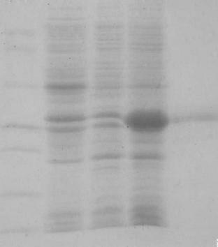
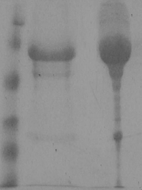
[0042] The identified positive recombinant plasmid pET-28a-Nsp9 was transformed into BL21(DE3) competent cells, and the bacteria were picked on the plate (Kan100 mg / mL), and the positive bacteria were coated with kanamycin (100 mg / mL) in LB liquid medium (Kan 100 mg / mL) overnight, then inoculated at 1% in LB medium (Kan 100 mg / mL) containing kanamycin, and cultured at 37°C until the logarithmic growth phase (OD600nm= 0.6), add IPTG (final concentration 1mmol / L), and induce culture at 37°C for 3 h. Centrifuge and precipitate the bacteria, and after ultrasonic crushing, obtain high-purity recombinant protein by electroelution, and store it at -80°C for future use. For identification results, see figure 1 .

Embodiment 2

[0043] Example 2 Preparation of Anti-Porcine Reproductive and Respiratory Syndrome Virus Nsp9 Protein Monoclonal Antibody

[0044] 1. For animal immunization, select healthy Balb / C mice aged 6-8 weeks to emulsify the purified PRRSV Nsp9 protein with complete Freund's adjuvant, and inject about 100 μg intraperitoneally into each mouse, and emulsify the protein intraperitoneally with incomplete Freund's adjuvant 14 days later. 100μg was injected, and at the last booster immunization, 100μg purified protein was directly injected intraperitoneally, and 50μg purified protein was injected into tail vein 3-4 days before fusion.

[0045] 2. Cell fusion Take splenocytes from immunized mice and mix them with SP2 / 0 in a fusion tube, centrifuge at 300g for 10 min, discard the supernatant, shake the cells to mix the two cells as evenly as possible, and then slowly drop and preheat within 60 seconds PEG-4000 solution, then slowly add serum-free 1640 medium to terminate the fusion, let it ...

Embodiment 3

[0054] Example 3 Establishment of blocking ELISA method

[0055] 1. The establishment of the reaction program

[0056] (1) Procedure of indirect ELISA method:

[0057] Coating: Dilute the ELISA antigen diluted with pH9.6 carbonic acid buffer to 0.2 μg / well, add 100 μL per well to the microtiter plate, and leave overnight at 4°C. Dry the coating solution and wash the plate 3 times with PBST (PBS containing 0.05% Tween-20, pH7.2). Blocking: Add 5% skimmed milk blocking solution, 300 μL / well, 37°C for 60 minutes, spin dry, wash with washing solution 3 times, 300 μL / well, 3 minutes each time. Adding samples: add anti-PRRSV Nsp9 protein monoclonal antibody 2D6, 100 μL per well, and react at 37°C for 60 min; wash with washing solution 5 times, 300 μL / well, 3 min / time. Add HRP-labeled goat anti-mouse HRP enzyme-labeled secondary antibody, 100 μL per well, and react at 37°C for 45 min; shake off the liquid, wash the plate, add TMB, 100 μL per well, and react at room temperature ...


PUM


Abstract

The invention relates to an activated toxic antibody kit for detecting a porcine reproductive and respiratory syndrome virus, which is characterized by being implemented by comprising the following steps of: after diluting a serum sample diluent to be detected twice, adding 100 microliters into an ELISA (Enzyme-Linked Immuno Sorbent Assay) plate for hatching at 37 DEG C for 1 hour; setting positive control, negative control and blank control; cleaning for five times with washing liquid every two minutes; then adding 100 microliters into each combined monoclonal antibody hole, hatching at 37 DEG C for 1 hour and cleaning for five times with washing liquid every two minutes; then adding goat-antimouse IgG-HRP ligature for acting at 37 DEG C for 1 hour and cleaning for five times with washing liquid every two minutes; then adding a developing substrate solution; mixing the substrate A with the substrate B according to the proportion of 1:1; adding 100 microliters into the ELISA hole; developing for 15 minutes by keeping in dark place; and adding 50 microliters of stop solution and detecting the OD490 value. The judging result is set according to the principle that the sample corresponding to the inhibition ratio PI which is equal to (OD uninhibited-OD sample)/OD uninhibited*100 percent, PI) 30 percent is positive, and the sample smaller than 30 percent is negative. The kit method has mature technology and strong repeatability, can effectively distinguish porcine reproductive and respiratory syndrome virus active toxic and inactivated toxic antibody and can be finished just by a common researcher.

Description

technical field [0001] The invention relates to a live virus antibody kit for detecting porcine reproductive and respiratory syndrome virus, in particular to a liquid phase blocking ELISA preparation method capable of distinguishing live virus infection and inactivated virus immune pigs, belonging to a new Animal disease diagnostic reagents, used in the field of animal husbandry and veterinary medicine. The invention has far-reaching research and wide applicability, including epidemiological investigation of porcine reproductive and respiratory syndrome virus in the region, purification of pig farms and distinction of wild virus infection and inactivated virus immune pig herds. Background technique [0002] Porcine reproductive and respiratory syndrome (PRRS) is caused by porcine reproductive and respiratory syndrome virus (Porcine reproductive and respiratory syndrome virus. PRRSV) infection. Reproductive disorders and respiratory diseases in piglets and finishing pigs. P...

Claims


Application Information

Patent Timeline
Patent Type & Authority Applications(China)
IPC IPC(8): G01N33/577G01N33/569G01N33/531
Inventor 丁壮黄志强母连志吴娟杨闽楠张泉鹏
Owner JILIN UNIV
Why Eureka
Try Eureka