Preparation method of ascites tumor cell sensitized DC-CIK

A technology for DC-CIK and ascites tumors, applied in the biological field, can solve the problems of inability to present antigens, high price, unfavorable separation of tumor cells, etc.

Active Publication Date: 2012-01-18
广东省医学医疗有限公司
View PDF1 Cites 13 Cited by
  • Summary
  • Abstract
  • Description
  • Claims
  • Application Information

AI Technical Summary

Problems solved by technology

Since the specific antigens of many tumors have not been identified and the antigen mutations of tumors can resist the immune attack of a single antigen, there are certain limitations in sensitizing DC cells with specific antigens
In addition, the surface antigens of tumor cell lines may change during in vitro passage, and DC cells cannot present all the antigens of the tumor to effector T cells, which limits the anti-tumor effect of DC-CIK in vivo
[0007] There are many traditional methods for extracting tumor cells from ascites. Immunomagnetic bead method is more advanced, but requires specific tumor cell antibodies and is expensiv

Method used


Image

  • Preparation method of ascites tumor cell sensitized DC-CIK
  • Preparation method of ascites tumor cell sensitized DC-CIK
  • Preparation method of ascites tumor cell sensitized DC-CIK

Examples


preparation example Construction

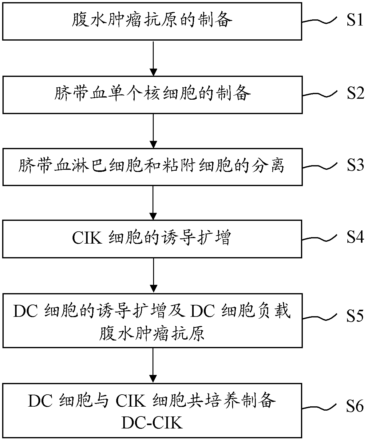
[0020] The embodiment of the present invention provides a method for preparing DC-CIK sensitized by ascites tumor cells capable of obtaining high proliferative activity and tumoricidal activity. The process flow of the preparation method of the ascites tumor cell sensitized DC-CIK is shown in Figure 1, including the following steps:

[0021] Step S1, preparation of ascites tumor antigen: resuspend ascites tumor cells in PBS buffer, then mix with leupeptin at a final concentration of 0.1-1.0 mg / ml to form a mixture, and place in liquid nitrogen for 5-10 minutes After that, put it in a water bath at 36-38°C. After the mixed solution is completely melted, put it in liquid nitrogen again, repeat 2-5 times, and then perform centrifugation in turn, and filter with a microporous membrane to obtain ascites tumor antigen;

[0022] Step S2, preparation of umbilical cord blood mononuclear cells: after centrifuging the obtained fresh umbilical cord blood, collect the upper layer of umbili...

Embodiment 1

[0043] The method for preparing DC-CIK sensitized by ascites tumor cells, as shown in Figure 1, includes the following steps:

[0044] S11. Preparation method of ascites tumor antigen:

[0045] (1) Isolate tumor cells from ascites: take 400ml of cancerous ascites, anticoagulate with 10IU / ml heparin sodium, mix the anticoagulated ascites with 6% hydroxyethyl starch at a ratio of 5:1, and statically Set aside for 1h, settle the red blood cells, take the supernatant, 600g, centrifuge for 5-10min, discard the supernatant, resuspend the cells with PBS, wash once, 600g, centrifuge for 5-10min, then resuspend the cells with 50ml PBS; take a 15ml centrifuge tube , respectively add 5ml of human tumor cell separation medium, density 1.055, carefully add 5ml of cell suspension to the top of the human tumor cell separation medium along the tube wall, be careful not to shake the centrifuge tube, centrifuge at 800g for 15min, and remove from the interface of human tumor cell liquid Tumor c...

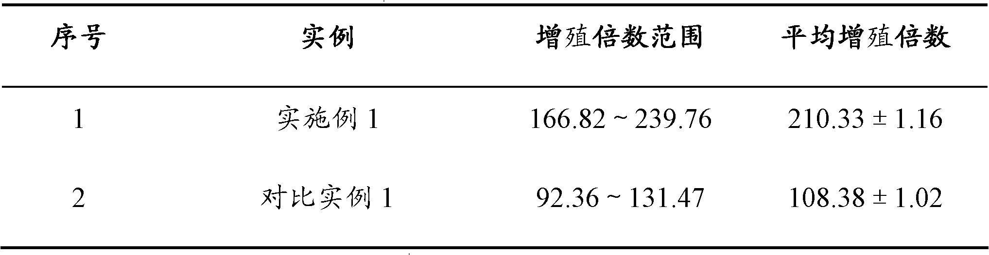
Embodiment 1 and comparative example 1

[0064] Example 1 and Comparative Example 1 Preparation of DC-CIK Cell Related Performance Index Detection

[0065] 1. Sterility and pyrogen testing:

[0066] It was found that the DC-CIK cells prepared in Example 1 and Comparative Example 1 were negative in both the sterility test and the heat source test.

[0067] 2. Phenotype detection:

[0068] The cell phenotype was detected by flow cytometry. The result of the phenotype of the DC-CIK cells prepared in Example 1 was: T lymphocytes (CD3+ cells) accounted for more than 98% of the total cells, and a small amount of DC. Among them, the proportion of T lymphocyte subsets has a certain range of variation due to individual differences: the proportion of CD3+CD56+ cells is 40-60%, and the proportion of CD3+CD8+ cells is 60-80%. The ratio of CD3+CD56+ cells in the DC-CIK prepared in Comparative Example 1 is 20-50%, and the ratio of CD3+CD8+ cells is 40-65%.

[0069] 3. Detection of cell proliferation activity:

[0070]

[00...


PUM

PropertyMeasurementUnit
Apertureaaaaaaaaaa

Abstract

The invention provides a preparation method of ascites tumor cell sensitized DC-CIK and application of the DC-CIK. The preparation method of ascites tumor cell sensitized DC-CIK comprises steps of: preparation of ascites tumour antigen; preparation of cord blood mononuclear cells; separation of cord blood lymphocytes and adherent cells; induction and amplification of the CIK cells; induction and amplification of the DC cells and loading of the ascites tumour antigen by the DC cells; and preparation of DC-CIK through co-culture of DC cells and CIK cells. The DC-CIK prepared by the preparation method of ascites tumor cell sensitized DC-CIK of the invention has high proliferative activity and tumor killing activity. The method has simple operations, easily controlled conditions and low equipment requirements.

Description

technical field [0001] The invention belongs to the field of biotechnology, and in particular relates to a method for preparing DC-CIK sensitized by ascites tumor cells and an application of DC-CIK. Background technique [0002] Immunotherapy is an important means of clinical treatment of tumors other than surgery, radiotherapy and chemotherapy. It plays an important role in removing residual and metastatic tumor cells in tumor patients, preventing tumor metastasis and recurrence, and improving patient survival and survival treatment. value. Currently, tumor immunotherapy methods mainly include lymphokine-activated killer cells (LAK), tumor-infiltrating lymphocytes (TIL), cytotoxic T lymphocytes (CTL), cytokine-induced killer cells (CIK), and natural killer cells ( NK), modified autologous dendritic cell-stimulated T lymphocyte therapy (DC-T), autologous dendritic cell-stimulated CIK immunotherapy (DC-CIK), etc. [0003] DC cells were first reported by Steinman and Cohn in...

Claims


Application Information

Patent Timeline
IPC IPC(8): C12N5/0783C12N5/0784
Inventor 刘韬
Owner 广东省医学医疗有限公司
Why Eureka
Try Eureka