Application of Raman encoding microsphere and method for detecting tumor marker by utilizing Raman encoding microsphere

A tumor marker and Raman coding technology, applied in the field of Raman coding microspheres, can solve the problems of harsh experimental conditions, complicated operation, and weak detection specificity of a single indicator, and achieve the effect of broadening the application scope.

Active Publication Date: 2012-01-18
HEFEI INSTITUTES OF PHYSICAL SCIENCE - CHINESE ACAD OF SCI
View PDF8 Cites 28 Cited by
  • Summary
  • Abstract
  • Description
  • Claims
  • Application Information

AI Technical Summary

Problems solved by technology

However, they all have disadvantages such as complicated operation, time-consuming and labor-intensive, and high cost to varying degrees.
For example, enzyme immunoassay technology requires special imported kits, and the acquisition of biological enzymes is difficult, the cost is high, and the experimental conditions are rela

Method used


Image

  • Application of Raman encoding microsphere and method for detecting tumor marker by utilizing Raman encoding microsphere
  • Application of Raman encoding microsphere and method for detecting tumor marker by utilizing Raman encoding microsphere
  • Application of Raman encoding microsphere and method for detecting tumor marker by utilizing Raman encoding microsphere

Examples


Embodiment 1

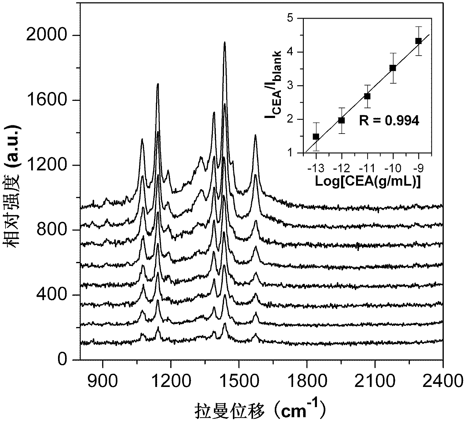
[0033] In this example, taking the tumor marker CEA as an example, the method for detecting the tumor marker CEA by using Raman-encoded microspheres is described.

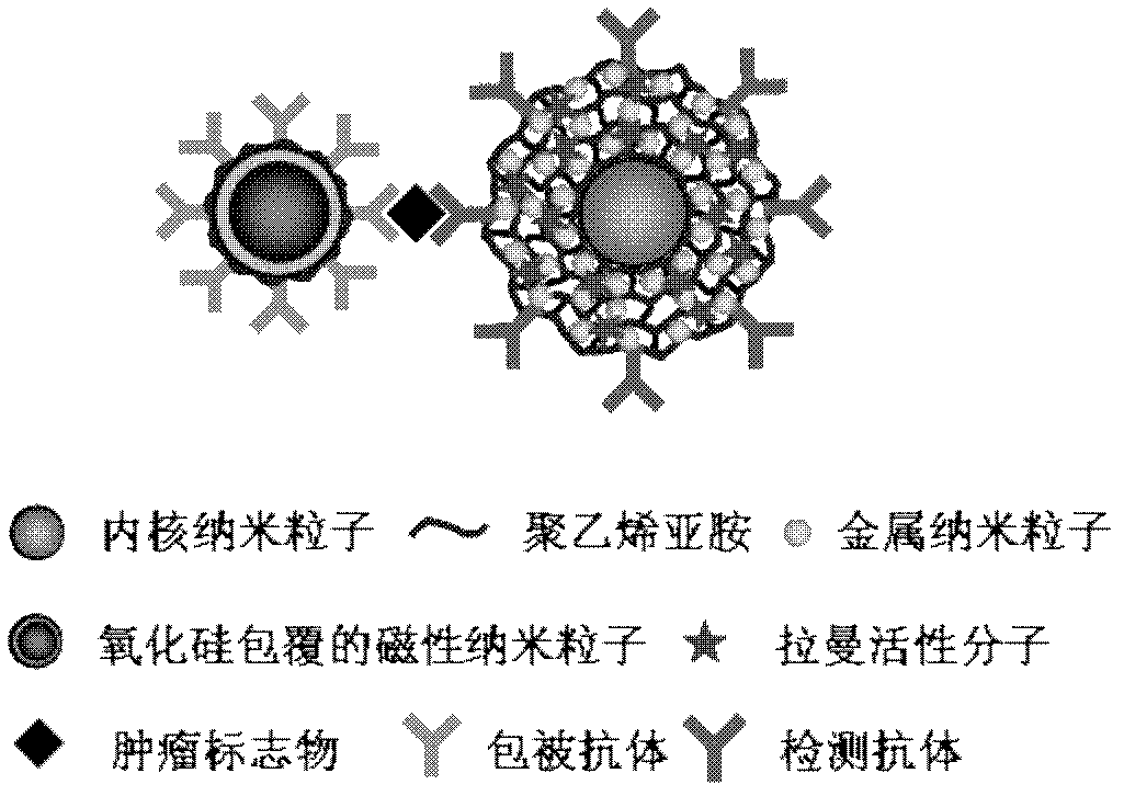
[0034] (1) Preparation of Raman-encoded microspheres

[0035] a. Take 95mL ultrapure water, add 1mL 30mM sodium citrate solution and 2mL 5mM silver nitrate solution in turn, then quickly inject 1mL 50mM sodium borohydride solution, stir at room temperature for 30 seconds, add 1mL 5mg / mL polyvinylpyrrolidone, the solution Gradually turn into deep yellow, and the silver nanoparticle sol with a diameter of 5-15nm is obtained.

[0036]b. Mix 3.6mL tetraethyl orthosilicate and 88.1mL ethanol into a 250mL round-bottomed flask, quickly add 11.9mL ammonia water under stirring conditions, and make it completely react at room temperature to obtain carbon dioxide with a diameter of about 200nm. Silicon nanoparticles.

[0037] c. Take 25mL of the silica nanoparticles prepared in step b, centrifuge and disperse in 50mL of 2mg...

Embodiment 2

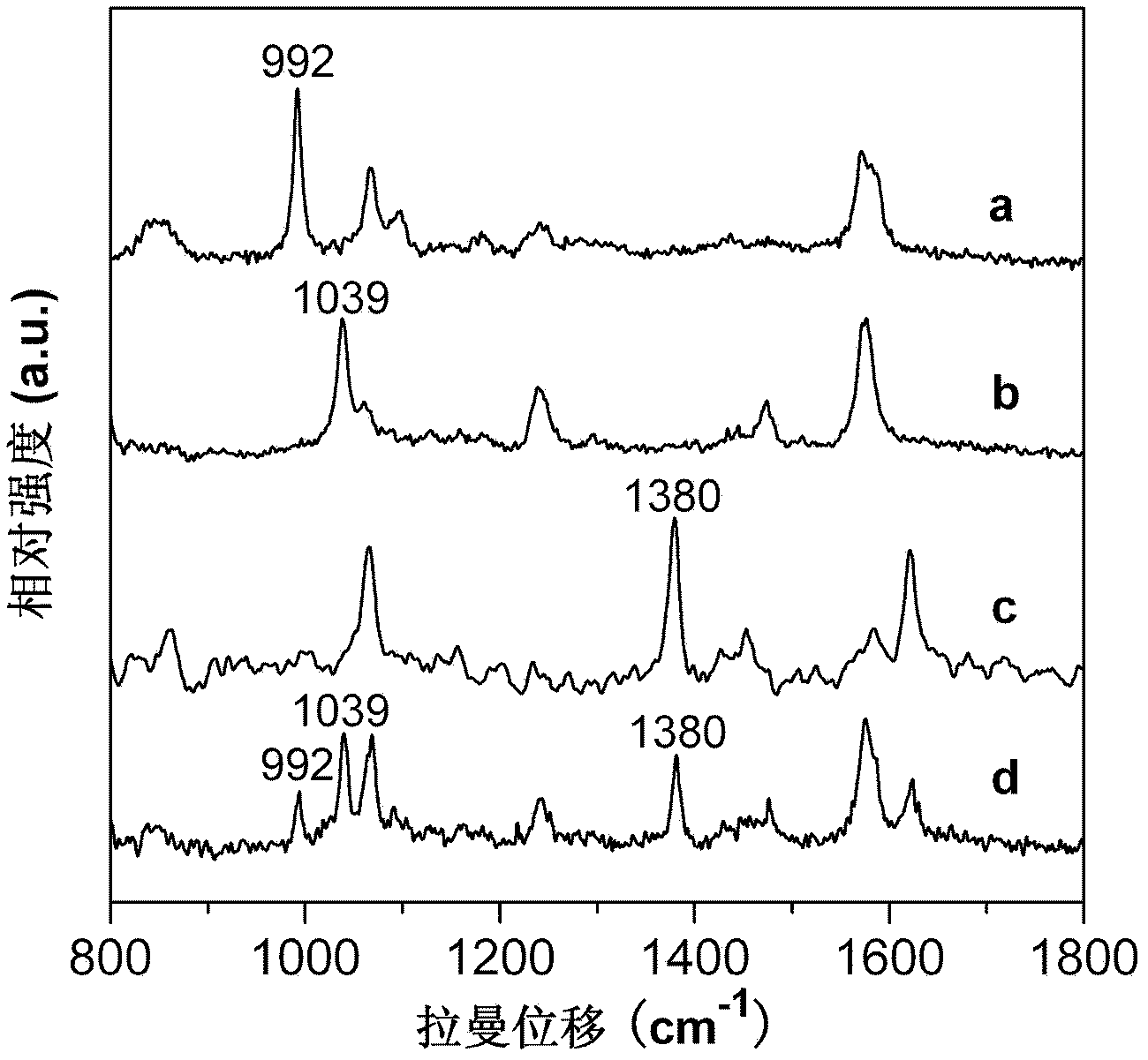
[0048] Example 2: Simultaneous detection of tumor markers AFP, PSA and CA125 by a Raman-encoded microsphere

[0049] (1) Preparation of Raman-encoded microspheres

[0050] The preparation method of the Raman coded microspheres in this example is the same as in Example 1, except that 3-methoxythiophenol, 2-methoxythiophenol and 2-naphthylthiophenol are used to replace p-mercaptoaniline as Raman active substances, respectively obtained encapsulated 3-methoxythiophenol-labeled core-shell nanospheres, encapsulated 2-methoxythiophenol-labeled core-shell nanospheres and encapsulated 2-naphthylthiol Labeled core-shell nanospheres.

[0051] (2) Preparation of Raman-encoded labeled nanoprobes

[0052] Add 125 μg of AFP antibody to 25 mg of the above-prepared encapsulated 3-methoxythiophenol-labeled core-shell nanospheres, react at 4°C for 12 hours, and finally block unreacted aldehydes with a mass concentration of 1% BSA base vacancy, centrifuged and washed to obtain 3-methoxythioph...


PUM


Abstract

The invention discloses an application of a Raman encoding microsphere and a method for detecting a tumor marker by utilizing the Raman encoding microsphere, wherein the application of the Raman encoding microsphere refers to the application of the Raman encoding microsphere in tumor marker detection. The method for detecting the tumor marker by utilizing the Raman encoding microsphere comprises the following steps: dispersing the Raman encoding microsphere into a phosphoric acid buffer solution and adding a detection antibody for reaction, thus obtaining a nano probe; then utilizing bovine serum albumin to seal a space bit on the surface of the nano probe, thus obtaining a nano prober marked by Raman encoding; adding serum containing a tumor marker into a solid-phase antibody, reacting and adding the nano prober marked by Raman encoding, thus obtaining an immune complex; and after enriching the immune complex through an additionally added magnetic field, performing SERS (surface enhanced Raman scattering) spectrum detection. The Raman encoding microsphere used in the invention has an ultra-strong SERS effect, and can be used for performing quantitative analysis on a polycomponent ultratrace object, and selecting a large number of molecules with different SERS characteristic oscillation as markers to detect various matters to be detected simultaneously.

Description

1. Technical field [0001] The invention relates to the use of Raman coded microspheres and a method for detecting tumor markers using Raman coded microspheres. 2. Background technology [0002] Malignant tumor is one of the important diseases that endanger human health. There are more than 10 million new cases and more than 6 million deaths in the world every year. In recent years, with the efforts of medical scientists and biologists, human beings' understanding of themselves has entered a microscopic level of analysis, and thus they have gained more understanding of the mechanism of tumor occurrence and development. A series of changes related to cell carcinogenesis were observed at the DNA, RNA, protein, chromosome, and cell levels. These abnormal changes are essentially signs of different stages of cell carcinogenesis. Using tumor-associated antigens as tumor markers and detecting them by immunoassay technology is expected to achieve early diagnosis of tumors. [0003]...

Claims


Application Information

Patent Timeline
IPC IPC(8): G01N33/574G01N33/76G01N21/65
Inventor 张忠平刘仁勇蒋长龙刘变化关贵俭
Owner HEFEI INSTITUTES OF PHYSICAL SCIENCE - CHINESE ACAD OF SCI
Why Eureka
Try Eureka