Electrochemical method employing DNA as probe to detect environmental pollutant

A detection environment and electrochemical technology, applied in the fields of biochemical analysis and biosensing, can solve the problems of insignificant changes in electrical signals, reduced instrument sensitivity, low response signals, etc., to expand the detection range, reliable detection results, and simple equipment. Effect

Active Publication Date: 2012-03-14
DALIAN INST OF CHEM PHYSICS CHINESE ACAD OF SCI
View PDF2 Cites 9 Cited by
  • Summary
  • Abstract
  • Description
  • Claims
  • Application Information

AI Technical Summary

Problems solved by technology

[0006] However, the response signal generated by the direct detection of DNA by electrochemical methods is low, and the weak response signal is easily interfered by noise, resulting in baseline shift, which reduces the sensitivity of the instrument.
However, the concentration of genotoxic compounds in the environment is low, and the electrical signal changes are not obvious

Method used


Image

  • Electrochemical method employing DNA as probe to detect environmental pollutant
  • Electrochemical method employing DNA as probe to detect environmental pollutant
  • Electrochemical method employing DNA as probe to detect environmental pollutant

Examples


Embodiment 1

[0042] Example 1. Nucleic acid biosensor assembly

[0043] Gold electrode assembly steps:

[0044] 1) The surface area is 0.02cm 2 The gold electrodes were polished with Al2O3 powders with particle sizes of 1, 0.3, and 0.05 μm, and then ultrasonically cleaned three times in absolute ethanol and deionized water for 2 minutes each time. Put it into 1M sulfuric acid solution and scan and activate it cyclically between 0 and 1.7V. When the scanning curves are completely overlapped, stop scanning (activation is complete).

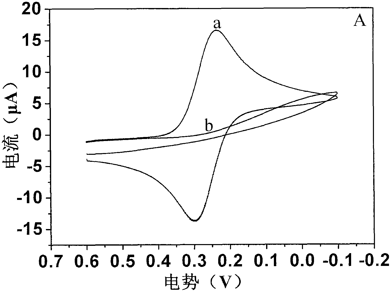
[0045] 2) figure 2 The cyclic voltammetry curve a in A indicates that the activated gold electrode is placed in 2mM potassium ferricyanide buffer solution, and the cyclic voltammetry curve is scanned between -0.1V and 0.6V. The curve redox peak potential difference is less than 75mV, indicating that the potassium ferricyanide reaction on the surface of the gold electrode is a completely reversible reaction, and the surface of the gold electrode has no impuri...

Embodiment 2

[0048] Embodiment 2. Nucleic acid biosensor detects hexachlorobenzene

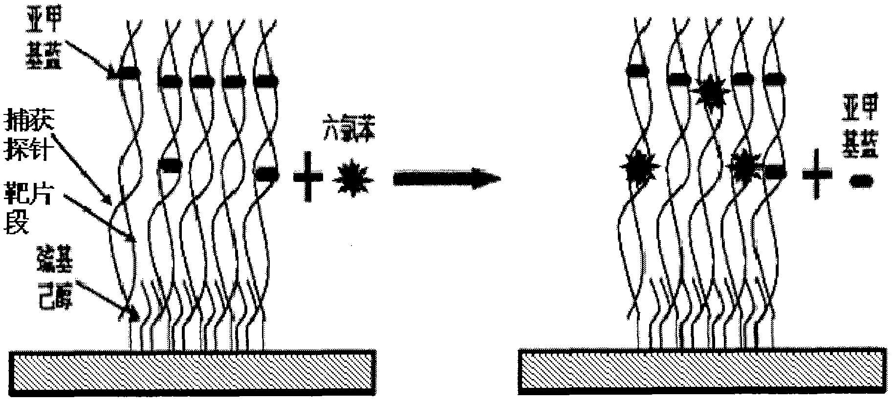
[0049] Hexachlorobenzene was dissolved in 10 mM PBS buffer solution with N, N-dimethylformamide as co-solvent to prepare 100 pM, 1 nM, 10 nM, 100 nM and 1 μM hexachlorobenzene solutions.

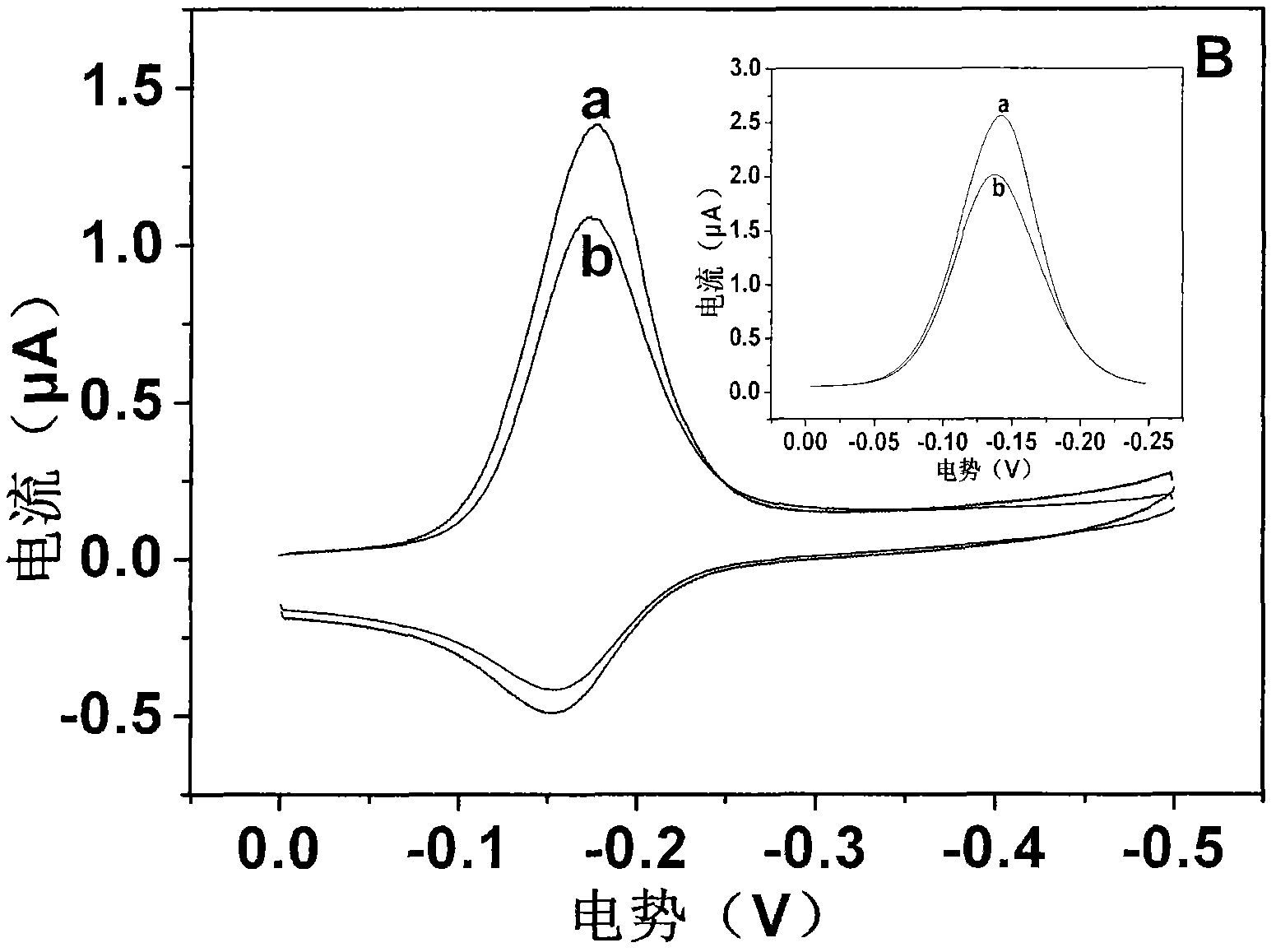
[0050] Figure 4 Representation: Add 15 μl of hexachlorobenzene solution dropwise to the surface of double-stranded DNA-modified gold electrode, treat it at room temperature for 20 minutes, then immerse in 10 μM MB of PBS (10 mM) buffer solution and scan electrochemically under nitrogen atmosphere to obtain the relationship between the concentration and the electrical signal related curves. Hexachlorobenzene interacts with DNA, causing changes in DNA pairing through non-covalent binding to double-stranded DNA, DNA base damage, etc., resulting in changes in the electrical signal of the nucleic acid sensor. As the concentration increases, the electrical signal gradually decays, and the intensity of the signal change is l...

Embodiment 3

[0051] Example 3. Ultraviolet-visible detection of the mode of action of hexachlorobenzene and DNA

[0052] Figure 5 Indicates: add 2ml of 10mM PBS buffer solution to the quartz cuvette, scan the blank at full wavelength within the range of 190-700nm, curve a is adding 20μl of 1mM MB to the blank solution, and scan at full wavelength to get the MB peak. Curve b is the mixed peak of MB and DNA obtained by adding 6 μl of 100 μM double-stranded DNA to the MB solution, shaking well and standing for 10 minutes. Select the non-interference peak at 620-700nm as the detection wavelength range. Curve c is adding 2μl 10 to the mixed solution of MB and DNA -2 M hexachlorobenzene, measure the absorption peak of the solution at 620-700nm.

[0053] According to the analysis of the experimental results, the MB peak appears at 190-350nm and 620-700nm, while the DNA peak is at 220-270nm, which interferes with the MB 190-350nm peak, so it is not suitable for analysis and research. The peak...


PUM


Abstract

An electrochemical method employing DNA as a probe to detect environmental pollutants. A biosensor detector comprises a working electrode, an auxiliary electrode, a reference electrode, a detection cell and an electrochemical working station. The working electrode is modified with nucleic acid probes on a surface thereof, and a genotoxicity compound in an environment sample is detected in a buffer system. When a detection result is positive, a signal of the sensor is changed obviously. Compared with an existing biosensor, the method has main advantages of: 1. high sensitivity; combination of an electrical active molecule and nucleic acid probes to increase a response current; 2. miniature and cheap apparatus and equipment, large detection flux, simple sample pre-treatment, and suitabilityfor on spot rapid screening detection. The method can not only carry out screening detection on a known genotoxicity compound but also evaluate potential toxicity effect of a newly synthesized compound; and the method can screen whether an actual sample contains a genotoxicity compound.

Description

technical field [0001] The invention belongs to the fields of biochemical analysis and biosensing, in particular to a method for detecting genotoxic compounds in the environment by a DNA electrochemical sensor. Background technique [0002] DNA sensor detection is mainly divided into target DNA detection and DNA mutation detection. These assays have broad applications in human disease diagnosis, pathogen detection in the environment, food safety, quarantine, etc. [0003] As people pay more and more attention to environmental safety and physical health, there is an urgent need for early diagnosis and treatment of various major diseases, and rapid safety assessment of the surrounding environment. However, the current screening and detection of genotoxic substances in the environment mainly obtains the data of their toxicity through animal experiments and cytotoxicological evaluation. This method is expensive and takes a long time, and is not suitable for the toxicity screeni...

Claims


Application Information

Patent Timeline
Patent Type & Authority Applications(China)
IPC IPC(8): G01N27/49G01N27/30
Inventor 吴立冬卢宪波苏凡陈吉平
Owner DALIAN INST OF CHEM PHYSICS CHINESE ACAD OF SCI
Why Eureka
Try Eureka