Method for separating mesenchymal stem cells from placenta

A technique for stem cells and placenta, applied in the field of stem cell isolation

Active Publication Date: 2012-09-19
BOYALIFE
View PDF8 Cites 47 Cited by
  • Summary
  • Abstract
  • Description
  • Claims
  • Application Information

AI Technical Summary

Problems solved by technology

[0006] The purpose of the present invention is to solve the defects of existing methods for obtaining placental mesenchymal stem cells, and provide a practical and simple method for isolating a large number of mesenchymal stem cells from placenta and optionally establishing a placental stem cell bank

Method used


Image

  • Method for separating mesenchymal stem cells from placenta
  • Method for separating mesenchymal stem cells from placenta
  • Method for separating mesenchymal stem cells from placenta

Examples


Embodiment 1

[0058] Embodiment 1, the separation of placental MSC

[0059] Within four hours after delivery, the placenta leaflets were cut off under aseptic conditions, and the placenta leaflets were fully washed with PBS buffer solution containing 10% volume FBS to remove residual blood in the placenta leaflets. First cut the placenta leaflet into 1cm 3 Tissue pieces of the same size were added to PBS buffer containing 0.1mg / mL dispase, 0.25mg / mL trypsin, 0.25mg / mL DNase I, 1mg / mL collagenase IV, 1mg / mL hyaluronidase for digestion at 37°C After 15 minutes, an appropriate amount of FBS was added to terminate the digestion. Then filter the tissue block and cell suspension together with a 200-mesh copper mesh and grind the tissue block with a syringe plunger, collect the filtered cell suspension and add it to a centrifuge tube for 10 minutes at 1000rpm, pour off the supernatant, and wash with 10% FBS Resuspend cells in PBS buffer. Then use density gradient centrifugation to separate an...

Embodiment 2

[0060] Embodiment 2, subculture of placental MSC and cryopreservation thereof

[0061] After about 10 days, after the scattered adherent cells formed colonies, they were digested with 0.05% trypsin / 2mMEDTA, and counted at 3000 cells / cm 2 Carry out subculture. Afterwards, subculture was digested when the cells reached about 70% confluency. Take 3×10 after digestion 6 The cells were added to 1ml of cell freezing solution (containing 50% low-sugar DMEM culture medium, 40% FBS, 10% dimethyl sulfoxide), and were cooled by a program, and finally entered into a liquid nitrogen tube for freezing.

Embodiment 3

[0062] Embodiment 3, biological characteristic identification of placental MSC

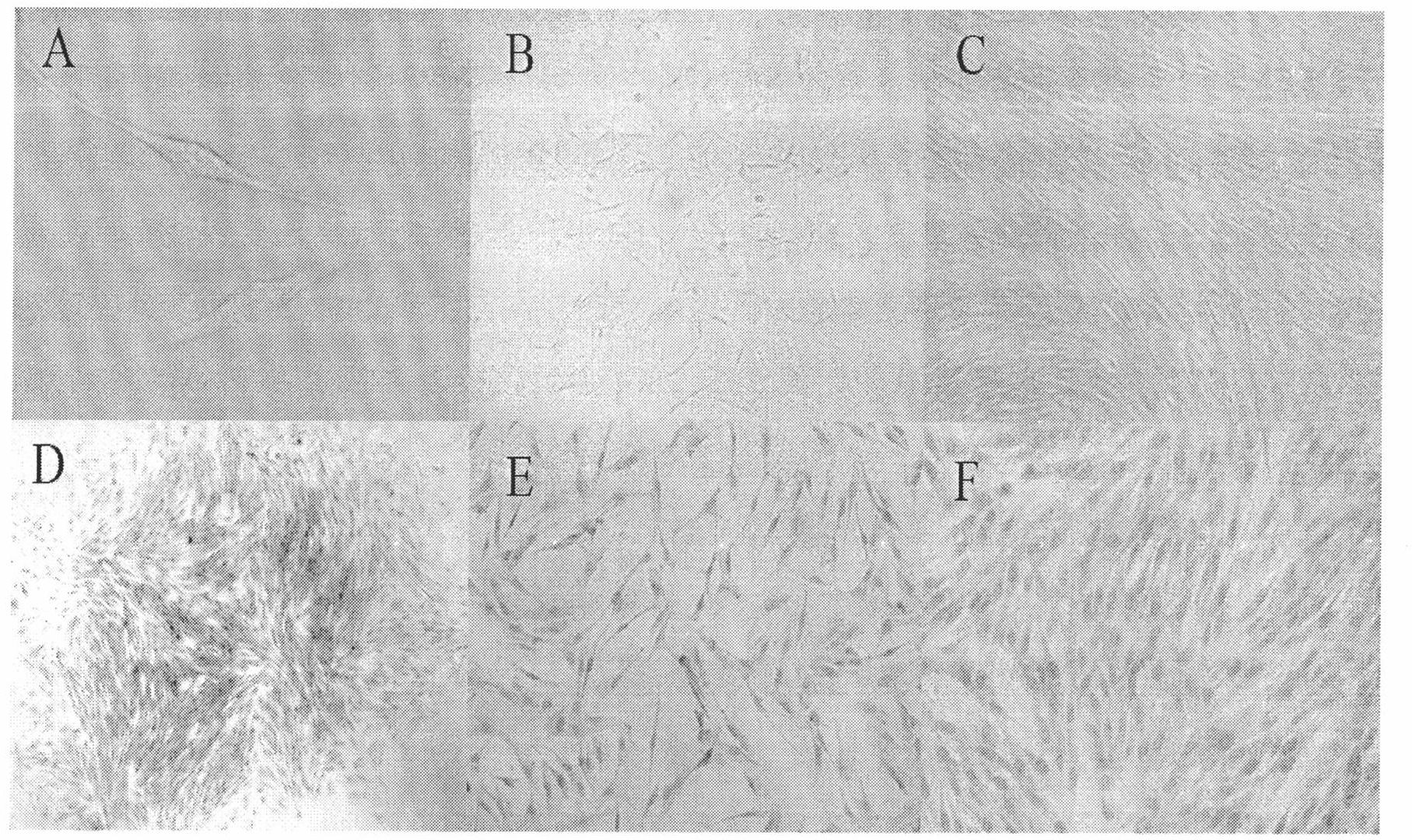
[0063] 1. Cell growth and morphological characteristics

[0064] By the isolation culture of embodiment 1 and embodiment 2, the placental mononuclear cells can be clearly seen under the microscope after the placental mononuclear cells are cultured for 72 hours. Spindle-shaped adherent cells ( figure 1 A), about 10 days will form turbine-shaped cell clones ( figure 1 B. figure 1 D), after digestion and passage, about 80% of the fusion adherent layer will be formed ( figure 1 C. figure 1 E. figure 1 F). During the culture process, it was found that the cell shape was relatively uniform, the proliferation speed was fast, the adhesion speed was fast, and it was easily digested by trypsin. After passage to more than 15 generations, its shape and growth characteristics did not change significantly.

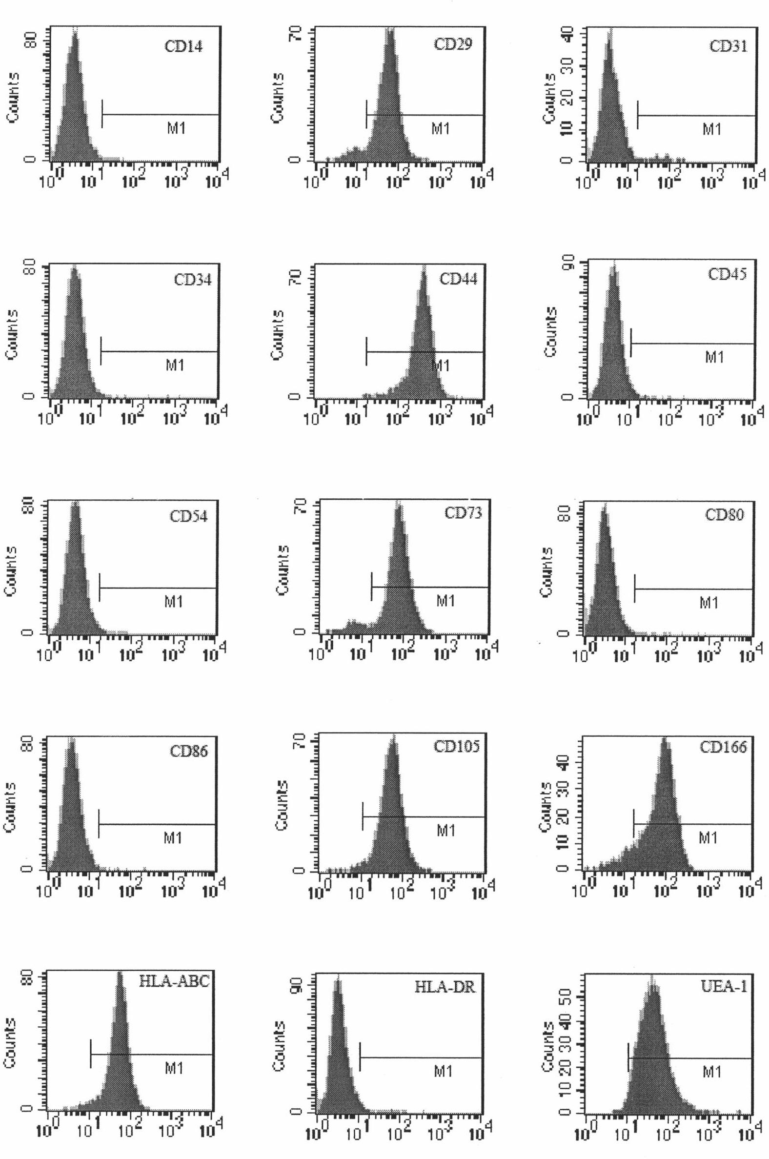
[0065] 2. Identification of MSC surface markers by flow cytometry

[0066] The 3rd, 6th, 9t...


PUM


Abstract

The invention relates to a method for separating mesenchymal stem cells from placenta. The method comprises the following steps: (a) taking placental cotyledon, and fully washing by using a phosphate buffer solution (PBS) to remove residual blood from the placenta; (b) cutting the placental cotyledon into blocks, adding a PBS containing tissue digestive enzyme, and incubating and digesting at 37DEG C; (c) filtering the tissue blocks by using a copper net, and grinding if necessary to promote filtration; (d) centrifuging the collected filtrate, separating mononuclear cells, suspending the obtained cells by using a mesenchymal stem cell (MSC) culture medium, and culturing in a 5 percent CO2 incubator at 37DEG C; and (e) after the dispersed cells form clones, selecting the clone cells, respectively culturing by using an MSC culture medium, and after the cells are fused, performing digestion and passage by using pancreatin to obtain the mesenchymal stem cells of the placenta. By the method, high purity mesenchymal stem cells of the placenta can be obtained.

Description

technical field [0001] The present invention relates to a method for isolating stem cells from placenta, in particular to a method for isolating mesenchymal stem cells from placenta. Background technique [0002] Mesenchymal stem cells (mesenchymal stem cells, MSCs) such as human mesenchymal stem cells were first isolated from bone marrow, a type of tissue stem cells derived from mesoderm with multi-lineage differentiation potential and self-renewal ability, in vivo and Under specific conditions in vitro, it has the ability to differentiate into various adult cells such as osteoblasts, chondrocytes, adipocytes, endothelial cells, nerve cells, muscle cells, and liver cells (Caplan AI. Mesenchymal stem cells. J Orthop Res. 1991, 9 : 641-650. Pittenger MF, Mackay AM, Beck SC, et al. Multilineage potential of adult human mesenchymal stem cells. Science. 1999; 284: 143-147). The latest research shows that mesenchymal stem cells have immune regulation and hematopoietic support fu...

Claims


Application Information

Patent Timeline
Patent Type & Authority Applications(China)
IPC IPC(8): C12N5/0775C12N5/0735G06F19/28
Inventor 霍思维陈俊峯张毅许晓椿李诣书
Owner BOYALIFE
Why Eureka
Try Eureka