Method for detecting regulatory T lymphocytes of human nasal mucosa

A technology for lymphocytes and nasal mucosa, applied in the field of biomedicine, can solve the problems such as the inability to directly react the Foxp3 protein of Treg cells, the inability to directly react to Treg cells, the complicated technical steps, etc., and achieve simple acquisition, simple and fast sample processing, and simple determination methods. Easy to operate effect

Inactive Publication Date: 2014-06-18
SHANGHAI PUTUO DISTRICT PEOPLES HOSPITAL
View PDF1 Cites 0 Cited by
  • Summary
  • Abstract
  • Description
  • Claims
  • Application Information

AI Technical Summary

Problems solved by technology

This is only an indirect detection method and cannot directly reflect the number of Treg cells and the expression of Foxp3 protein
[0006] 3. The tissue is digested with collagenase to obtain single cells, and then the expression of Treg cells is detected by flow cytometry. Treg cells can be analyzed by detecting a large number of cells, but the technical steps are cumbersome, and the tissue needs to be cut into 2mm first. 3 Size, pre-digestion solution at 37°C for 30min, centrifugation, collagenase digestion for 60min, and cell filtration
The existing technology uses RT-PCR to detect the expression of Foxp3 gene, which is only an indirect detection method and cannot directly reflect the number of Treg cells and the expression of Foxp3 protein

Method used


Image

  • Method for detecting regulatory T lymphocytes of human nasal mucosa
  • Method for detecting regulatory T lymphocytes of human nasal mucosa
  • Method for detecting regulatory T lymphocytes of human nasal mucosa

Examples


preparation example Construction

[0062] (2) Preparation of single-cell suspension: Put the nasal mucosa into 1ml of normal saline, and blow gently with a pipette to disperse the cells evenly. Centrifuge at 5000rpm for 10 minutes, discard the supernatant, resuspend the pellet with 200ul PBS, and count.

[0063] (3) Regulatory T cell (Foxp3) detection (BD Pharmingen, CA, USA)

[0064] Reagent preparation: Buffer A and Buffer C are freshly prepared before use

[0065] 1× Human Foxp3 Buffer A: Dilute 10× Human Foxp3 Buffer A with deionized water to 1× working concentration.

[0066] Preparation of Working Solution Human Foxp3 Buffer C: Mix Foxp3 Buffer B with 1×Human Foxp3 Buffer A at a ratio of 1:50.

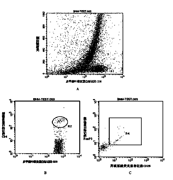
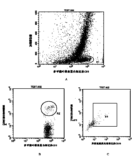
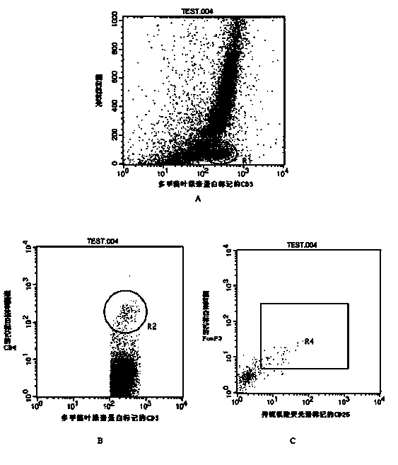
[0067] 1) Surface staining: Add 100ul cells (about 1×10⁵ cells) to a 12×75 mm flow tube, and add cell surface staining antibodies.

[0068] Isotype control tube: CD3-PerCP 5ul + CD4-APC 2ul + IgG1-FITC 5ul

[0069] Test tube: CD3-PerCP 5ul + CD4-APC 2ul + CD25-FITC 5ul; Vortex the two tubes at room tempe...

Embodiment

[0078] Detection of Treg cells in nasal mucosa of patients with allergic rhinitis and normal people.

[0079] (1) Diagnosis and source of patients with allergic rhinitis

[0080] Allergic rhinitis patients were from outpatients of our hospital, and the diagnosis was in accordance with the "Diagnostic Criteria and Treatment Guidelines for Allergic Rhinitis" formulated by the Otolaryngology-Head and Neck Surgery Branch of the Chinese Medical Association. Exclusions included patients with chronic rhinosinusitis and nasal polyps, severe deviated nasal septum, and other chronic heart, lung, and endocrine dysfunction. Normal people come from the medical examiners of our hospital.

[0081] (2) Detection of Treg cells

[0082] Use a nasal mucosal curette to scrape off the nasal mucosa at the lower and middle end of the lower turbinate, place in normal saline, blow and mix well, centrifuge, discard the supernatant, obtain cell pellets, and count. Label CD3, CD4, and CD25 on the cell...


PUM


Abstract

The invention belongs to the field of biotechnology, and discloses a method for detecting regulatory T lymphocytes (Treg) of human nasal mucosa. The method comprises the following steps: 1) obtaining nasal mucosa cells: enabling a spoon-shaped tail end of nasal mucosa curette to extend into a nasal cavity to the middle and lower end of inferior turbinate, lightly pressing the curette on the surface of the nasal mucosa, then moving outwards for 4-5mm, repeating the action for 2-3 times, taking out the curette, observing white sticky substance in the spoon tail end and obtaining a nasal mucosa specimen; 2) preparing monoplast suspension liquid: putting the nasal mucosa specimen in normal saline, and lightly beating to enable the cells to be uniformly dispersed so as to obtain the monoplast suspension liquid; 3) detecting the regulatory T lymphocytes: analyzing the T lymphocytes of the monoplast suspension liquid by flow cytometry.

Description

technical field [0001] The invention belongs to the technical field of biomedicine and relates to cellular immunology. More specifically, the invention relates to a method for detecting regulatory T lymphocytes of human nasal mucosa. Background technique [0002] The nasal mucosa is a part of the upper respiratory tract mucosa and is the first battlefield where inhaled antigens, toxins and microorganisms interact with the body's immune system. The defense mechanism of nasal mucosa is regulated by humoral and cellular immune responses, involving immunocompetent cells including T and B lymphocytes. Allergic rhinitis (AR) is a group of local Th2 cytokines (IL-4, IL-5, IL-9, IL-13) in the nasal cavity caused by stimulation by environmental antigens such as dust mites, plant pollen, and animal protein. A disease characterized by predominant expression, selective infiltration of eosinophils, mucosal hyperreactivity, and excessive IgE synthesis. Among them, the reduction of regul...

Claims


Application Information

Patent Timeline
Patent Type & Authority Patents(China)
IPC IPC(8): G01N15/14
Inventor 李文秀白素娟金姝闫佩毅庞权
Owner SHANGHAI PUTUO DISTRICT PEOPLES HOSPITAL
Why Eureka
Try Eureka