
Cell model and method for screening c-Fms kinase inhibitor

A cell and cell line technology, applied in the fields of cell biology and pharmacy, can solve the problems of cumbersome operation steps, low degree of quantification, and difficulty in accurate detection, and achieve the effect of simple, fast, and high sensitivity

Inactive Publication Date: 2012-10-10
INST OF PHARMACOLOGY & TOXICOLOGY ACAD OF MILITARY MEDICAL SCI P L A
View PDF1 Cites 2 Cited by
  • Summary
  • Abstract
  • Description
  • Claims
  • Application Information

AI Technical Summary

Problems solved by technology

[0005] According to current literature reports, the methods for screening c-Fms inhibitors mainly include molecular level kinase activity assay and cellular level kinase phosphorylation immunoblotting detection method, but there are disadvantages such as cumbersome operation steps and low degree of quantification
In addition, because the phosphorylation of receptor kinases such as c-Fms is a short-term process, it is difficult to detect accurately, and it is prone to dimerization in vitro. So far, there has been no high-content analysis and high-throughput screening of c-Fms. Report on Cell Model of Fms Inhibitor

Method used


Image

  • Cell model and method for screening c-Fms kinase inhibitor
  • Cell model and method for screening c-Fms kinase inhibitor
  • Cell model and method for screening c-Fms kinase inhibitor

Examples


Embodiment 1

[0059] Example 1: Establishment of cell lines stably expressing human c-Fms and STAT1

[0060] Construct the pCORON / puro-cfms eukaryotic expression vector containing the full-length cDNA of human c-Fms (gifted by Dr. Charles Sherr and Dr. Martine Roussel, St. Jude Children's Research Hospital, USA), and liposome transfection has been fused to express GFP and STAT1 human osteosarcoma cell line.

[0061] A novel cell line stably expressing human c-Fms was obtained by resistance screening and limiting dilution. Use DMEM medium containing 4500 mg / L glucose, 10% fetal bovine serum, 4 mM L-glutamine, 500 μg / mL G418 and 2 μg / mL puromycin at 37 °C, 5% CO 2 , cultivated in an 80% humidity incubator.

[0062] Compared with before transfection, the morphology of the obtained cells did not change significantly, and the growth state was stable.

[0063] The cell line prepared in this example (ie, the preserved human osteosarcoma cell line in the present invention, with the deposit num...

Embodiment 2

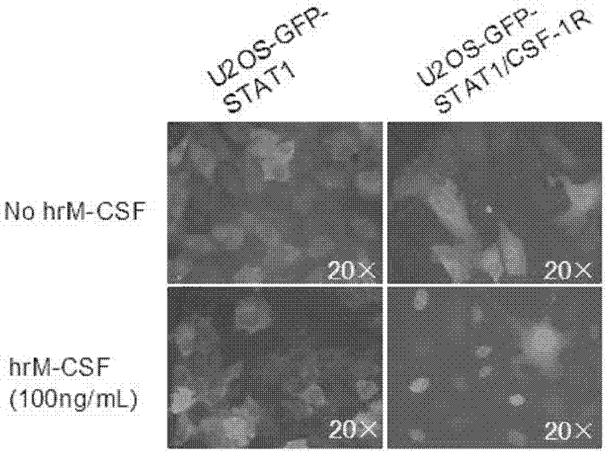
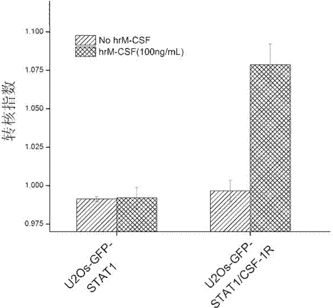
[0064] Example 2: Identification of newly established cell lines

[0065] The cell line prepared in Example 1 was treated with recombinantly expressed human M-CSF (hrM-CSF) to activate c-Fms and induce nuclear translocation of GFP-STAT1. After the human macrophage colony-stimulating factor receptor in the established cell line binds to its specific ligand M-CSF, the receptor dimerizes and the spatial conformation changes to activate the kinase domain. Through transphosphorylation, the signaling pathway downstream of c-Fms is activated. Phosphorylated GFP-STAT1 also forms dimerization, enters the nucleus, and undergoes nuclear translocation.

[0066] In order to quantitatively detect the degree of nuclear translocation of GFP-STAT1 induced by hrM-CSF, in this example, In Cell Analyzer 1000 or In Cell Analyzer 2000 was used to obtain cell images of GFP-STAT1 nuclear translocation, which were analyzed by the Nuclear Trafficking Analysis Module. The cell image of , the degree ...

Embodiment 3

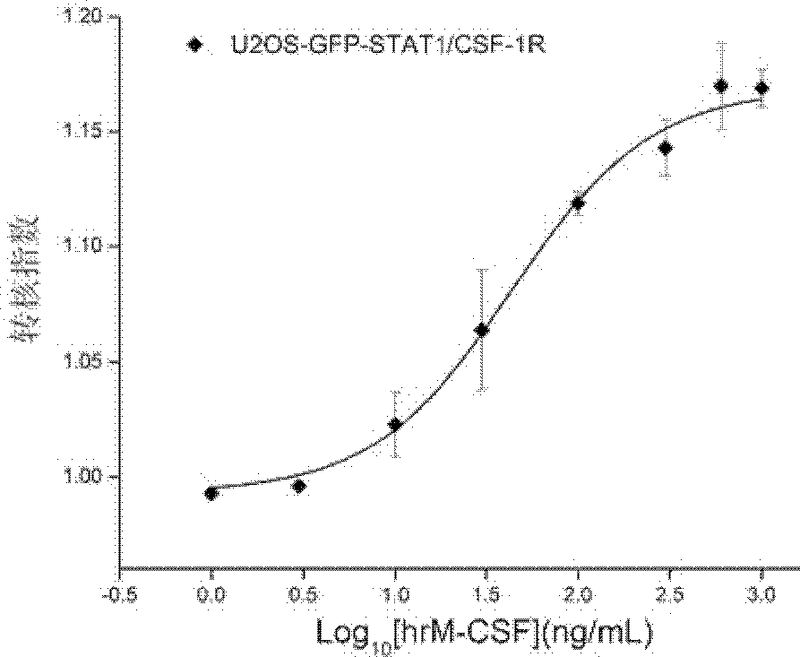
[0074] Example 3: Determination of the half effective concentration of hrM-CSF-induced GFP-STAT1 nuclear transfer

[0075] Proceed as follows:

[0076] 1) The cells prepared in Example 1 were made into 1×10 5 cells / mL of cell suspension, inoculated in a black transparent bottom 96-well culture plate, 100 μL / well.

[0077] 2) At 37°C, 5% CO 2 , cultivated in an 80% humidity incubator for 24 hours.

[0078] 3) Wash the cells twice with the cell analysis solution, 100 μL / well each time; discard the solution, and then add 50 μL / well of the cell analysis solution.

[0079] 4) Add hrM-CSF diluted in the cell analysis solution to make the final concentrations respectively 1, 3, 10, 30, 100, 300, 600, and 1000 ng / mL.

[0080] 5) 37°C, 5% CO 2 After incubating in an 80% humidity incubator for 30 minutes, add 37°C preheated cell fixation solution, 100 μL / well, shake the culture plate slightly to mix, and place it at room temperature in the dark for 20 minutes.

[0081]6) Discard...


PUM


Abstract

The invention belongs to the fields of cell biology and pharmacy and relates to a cell model and a method for screening a c-Fms kinase inhibitor, concretely to a cell which simultaneously expresses a macrophage colony stimulating factor receptor and an STAT1protein. The invention also relates to a method for screening a c-Fms tyrosine kinase inhibitor, a method for accessing inhibitory activity of a compound or a combination to the c-Fms tyrosine kinase and a purpose of the cell in screening the c-Fms tyrosine kinase inhibitor. The cell model established in the invention is sensitive, efficient, reliable and capable of being used in a high throughput screening and / or a high content screening of the c-Fms tyrosine kinase inhibitor.

Description

technical field [0001] The invention belongs to the field of cell biology and pharmacy, and relates to a cell model and a screening method for screening c-Fms kinase inhibitors. Background technique [0002] Drug screening models are an essential platform for drug activity research in drug development. High content analysis or high content screening (High Content Screening, HCS) HCS is an automated platform for high-throughput fluorescence / bright-field microscopy imaging and quantitative image analysis. Under the premise of completeness and functional integrity, simultaneously detect the effects of the screened samples on cell morphology, growth, differentiation, migration, apoptosis, metabolic pathways and signal transduction, obtain a large amount of relevant information in a single experiment, and determine its biological activity and potential toxicity. On the basis of high-content technology, the establishment of cell screening models for kinase inhibitors, combined w...

Claims


Application Information

Patent Timeline
Patent Type & Authority Applications(China)
IPC IPC(8): C12N5/10C12Q1/48C12Q1/02
CPCG01N2500/10C12Q1/485G01N33/5035C12Y207/01112C07K14/7153C07K14/4705
Inventor 王莉莉杨胜乾龙隆肖军海李松
Owner INST OF PHARMACOLOGY & TOXICOLOGY ACAD OF MILITARY MEDICAL SCI P L A
Learn More