Fluorescence in situ hybridization method of 5S rDNA on plant chromosome

A fluorescence in situ hybridization and chromosome technology, applied in the field of cytogenetics and molecular biology, to achieve the effect of simple method, simplified pretreatment steps, and simple elution steps

Active Publication Date: 2013-11-27
NANKAI UNIV
View PDF2 Cites 10 Cited by
  • Summary
  • Abstract
  • Description
  • Claims
  • Application Information

AI Technical Summary

Problems solved by technology

[0003] 5S rDNA probes are generally obtained by plasmid or PCR amplification, and then directly or indirectly labeled for FI...

Method used


Image

  • Fluorescence in situ hybridization method of 5S rDNA on plant chromosome
  • Fluorescence in situ hybridization method of 5S rDNA on plant chromosome
  • Fluorescence in situ hybridization method of 5S rDNA on plant chromosome

Examples


Embodiment 1

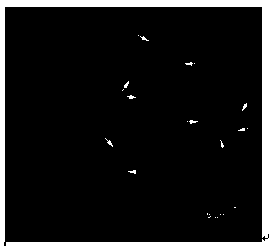
[0051] FISH Mapping of 5S rDNA on the Metaphase Chromosome

[0052] 1. Chromosomal Specimen Preparation

[0053] (1) The root tip of mustard mustard seeds grows to 0.5cm after germination.

[0054] (2) Cut off the root tip and pretreat it with saturated p-dichlorobenzene aqueous solution for 5 hours at room temperature.

[0055] (3) The root tip was hypotonic with 0.075mol / L potassium chloride hypotonic solution for 30 minutes, and then fixed with 3:1 (methanol: glacial acetic acid) for 20 minutes.

[0056] (4) Wash three times with distilled water, 5min each time, to fully wash away the fixative.

[0057] (5) Using 2.5% (w / w) pectinase and cellulase mixed in a weight ratio of 1:1, the root tip was dissociated at 37°C for 1 hour.

[0058] (6) Wash off the enzyme solution with distilled water, hypotonic with 0.075mol / L potassium chloride hypotonic solution for 30min, and fix with 3:1 methanol: glacial acetic acid fixative for 20min.

[0059] (7) Prepare chromosome specimens...

Embodiment 2

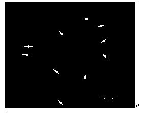
[0082] Embodiment 2: comparative embodiment

[0083] classic in situ hybridization

[0084] step:

[0085] Steps for FISH mapping of 5S rDNA on the metaphase chromosomes of potherb mustard:

[0086] 1. Chromosomal Specimen Preparation

[0087] (1) The root tip of mustard mustard seeds grows to 0.5cm after germination.

[0088] (2) Cut off the root tip and pretreat it with saturated p-dichlorobenzene aqueous solution for 5 hours at room temperature.

[0089] (3) The root tip was hypotonic with 0.075mol / L potassium chloride hypotonic solution for 30 minutes, and then fixed with 3:1 (methanol: glacial acetic acid) for 40 minutes.

[0090] (4) Wash three times with distilled water, 5min each time, to fully wash away the fixative.

[0091] (5) Using 2.5% (w / w) pectinase and cellulase mixed in a weight ratio of 1:1, the root tip was dissociated at 37°C for 1 hour.

[0092] (6) Wash off the enzyme solution with distilled water, hypotonic with 0.075mol / L potassium chloride hyp...

Embodiment 3

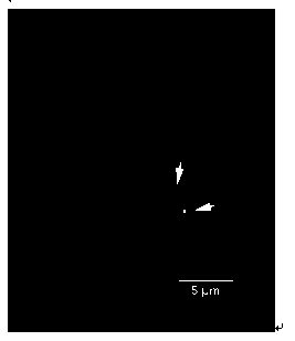
[0125] Examples of practical applications

[0126] Application method of the present invention has been in such as poplar (see image 3 ), cauliflower (see Figure 4 ),carrot( Figure 5 ) and more than 30 kinds of plants have been successfully tested on a variety of plants,

[0127] Method: with embodiment 1

[0128] Step: with embodiment 1

[0129]Conclusion: The method of the present invention can be extended and applied to 5S rDNA FISH localization of various plants.


PUM


Abstract

The invention discloses a fluorescence in situ hybridization method of 5S rDNA on a plant chromosome. The fluorescence in situ hybridization method mainly comprises the steps of chromosome specimen preparation, 5S rDNA probe preparation, chromosome fluorescence in situ hybridization, hybridization signal detection and the like. The invention mainly solves the problem that the labeling of the conventional fluorescence in situ hybridization probe is complicated and cumbersome and provides the simple, convenient, fast and accurate probe labeling and hybridization method. According to the fluorescence in situ hybridization method, the FISH positioning of the 5S rDNA on the plant chromosome can be completed within three hours. The 5S rDNA probe is an oligonucleotide probe. When the 5S rDNA probe is synthesized, fluorophore can be added into the 5S rDNA probe to modify the 5S rDNA probe, so that labeling is not needed. Compared with the prior art, the fluorescence in situ hybridization method is simple and low in cost.

Description

technical field [0001] The invention belongs to the technical field of cytogenetics and molecular biology, and relates to a method for fluorescent in situ hybridization of 5S rDNA on plant chromosomes. Background technique [0002] Ribosomal RNA gene (rDNA) is one of the most widely studied genetic units in the plant genome. rDNA is a family of highly conserved repetitive sequences with hundreds or even thousands of copies distributed in clusters on one or more pairs of chromosomes. The positioning of rDNA on chromosomes by fluorescence in situ hybridization (FISH) technology can provide effective cytological markers for karyotype analysis, especially for species with small chromosomes and similar shapes, this chromosome marker is more suitable for karyotype analysis. important tool. [0003] 5S rDNA probes are generally obtained by plasmid or PCR amplification, and then directly or indirectly labeled for FISH. There is no report on the use of fluorophore-labeled oligonucle...

Claims


Application Information

Patent Timeline
IPC IPC(8): C12Q1/68
Inventor 陈成彬王春国宋文芹
Owner NANKAI UNIV
Why Eureka
Try Eureka