HSV1-TK molecular imaging probe for detecting cell apoptosis as well as construction method and application thereof

A technology of HSV1-TK and molecular imaging, which is applied in the fields of biochemical equipment and methods, pharmaceutical formulations, microbial measurement/inspection, etc., and can solve the problems of low resolution, optical imaging penetration and tomographic resolution, application limitations, etc. question

Active Publication Date: 2014-06-04
XIDIAN UNIV
View PDF3 Cites 3 Cited by
  • Summary
  • Abstract
  • Description
  • Claims
  • Application Information

AI Technical Summary

Problems solved by technology

However, MRI and ultrasound imaging are limited due to their low sensitivity and optical imaging due to the...

Method used


Image

  • HSV1-TK molecular imaging probe for detecting cell apoptosis as well as construction method and application thereof
  • HSV1-TK molecular imaging probe for detecting cell apoptosis as well as construction method and application thereof
  • HSV1-TK molecular imaging probe for detecting cell apoptosis as well as construction method and application thereof

Examples


Embodiment 1

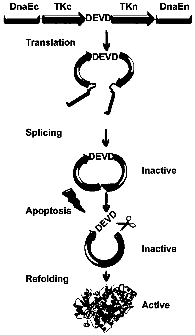
[0016] Embodiment 1 constructs HSV1-TK molecular image probe, figure 2 Shown:

[0017] 1. Linearized vector: pcDNA3.1 is digested with restriction endonucleases HindIII and XhoI, and the digested product is subjected to 1% agarose gel electrophoresis, and the target DNA fragment is excised under ultraviolet light. The target band was recovered and purified with a gel extraction kit.

[0018] 2. Get the insert fragment:

[0019] Obtaining the DEVD sequence: The DEVD sequence was synthesized from Shanghai Sangong Company using conventional chemical synthesis methods, and its sequence is 5'-gatgaagtcgac-3';

[0020] Obtaining TKn and TKc sequences: PCR amplified the N-terminal and C-terminal of HSV1-TK fragment respectively. In order to reduce the toxicity of TK in vivo, the first 45 amino acid sequences of its N-terminal were not amplified, but started from the 46th amino acid. After bioinformatics analysis, it is predicted that there are 3 potential sites, which can be int...

Embodiment 2

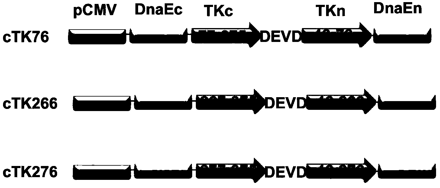
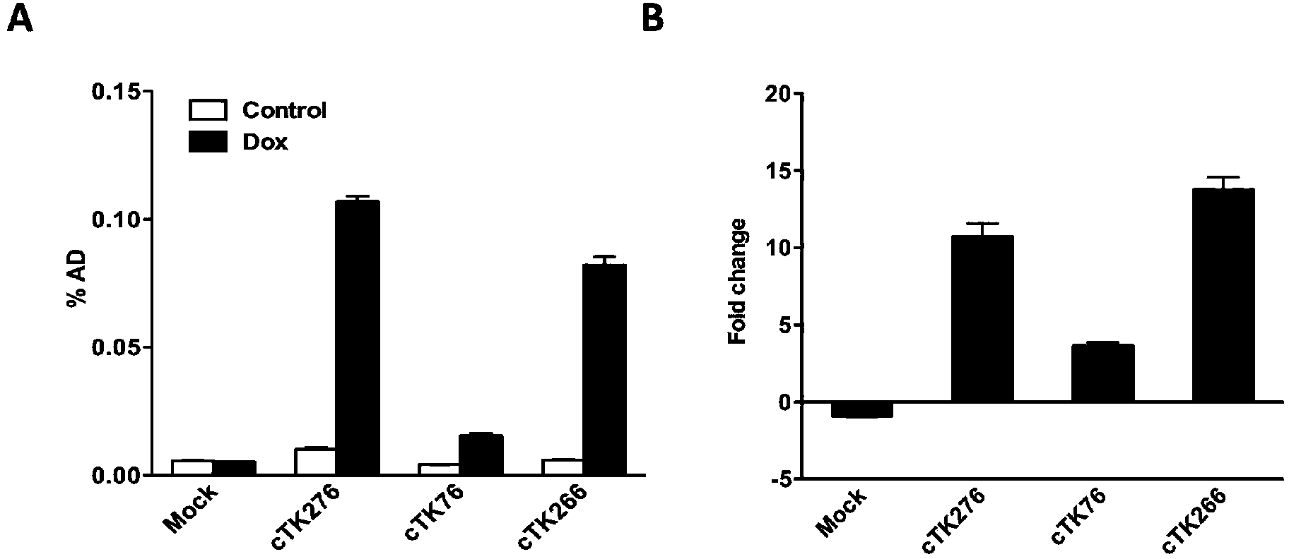
[0031] Example 2 Determination of Molecular Imaging Probe Activity During Apoptosis (see image 3 )

[0032] 1. To culture 22B tumor cells, take a 12-well culture dish and add a certain amount of 3x10⁵ 22B cells to culture at 37 degrees until about 70% confluence.

[0033] 2. Add 0.8ug of cTK76, cTK266 and cTK276 plasmid DNA respectively according to the instructions of Lipofectamine2000.

[0034] 3. After 24 hours of transfection, add 5ug / ml doxorubicin (Dox) to each well of cells to induce cell apoptosis.

[0035] 4. After 24 hours of induction, add 10uCi of radioactive substrate to each well of cells 18 After F-FHBG was cultured at 37 degrees for 1 hour, the culture medium was washed away, and then washed 3 times with PBS, and the cells in each well were lysed with 200ml of 0.1M NaOH.

[0036]5. Collect each well of cell lysate, calculate its radioactivity, expressed as % AD, and calculate the intracellular reaction of each probe 18 The relative change value of F-FHBG. ...

Embodiment 3

[0037] Determination of protein conformation of molecular imaging probes during apoptosis in Example 3 ( Figure 4 )

[0038] 1. To culture 22B tumor cells, take a 6-well culture dish, add a certain amount of 3x10⁶ 22B cells to 2 wells, and culture at 37 degrees to about 70% confluence.

[0039] 2. Add 1.6ug of cTK266 plasmid DNA according to the instructions of Lipofectamine2000.

[0040] 3. After 24 hours of transfection, add 5ug / ml doxorubicin (Dox) to one of the cells to induce apoptosis, and the other well without dox as a control.

[0041] 4. After 24 hours of induction, the cells were collected and detected by western blot with TK antibody. The result can be seen ( Figure 4 shown), when there is no dox induction (Dox is 0), the molecular probe is mainly cyclic TK band (cyclic TK) in the cell, but a slight linear TK band (linearTK) can also be seen, which means It is because the TK probe exists in the body mainly in the form of a ring-shaped protein structure before...


PUM


Abstract

The invention discloses an HSV1-TK (Herpes Simplex Virus 1-Thymidine Kinase) molecular imaging probe for detecting cell apoptosis as well as a construction method and application thereof. A preparation method of the HSV1-TK probe comprises the following steps: (1) performing enzyme digestion on a carrier pcDNA3.1 by using restriction enzymes HindIII and XhoI to linearize the carrier; (2) amplifying the terminal C of DnaE, the terminal C of a TK gene, a DEVD sequence, the terminal N of the TKgene and the terminal N of protein intron DnaE by virtue of PCR (Polymerase Chain Reaction); (3) recombining each segment with the linearized pcDNA3.1 carrier in a certain sequence by virtue of a gene recombination technology; (4) carrying out transformation, plasmid extraction and enzyme digestion identification on the recombinant carrier, thereby obtaining the HSV1-TK probe. The molecular imaging probe is capable of monitoring the occurrence and development of cell apoptosis in vivo, and thus overcoming the defect that the cell apoptosis only can be observed at an in-vitro level by virtue of technologies such as an electron microscope and flow cytometry and provides a favorable tool for monitoring in vivo.

Description

technical field [0001] The invention relates to the field of biotechnology, in particular to an HSV1-TK molecular imaging probe for detecting cell apoptosis and its construction method and application. Background technique [0002] Apoptosis is a physiological programmed cell death process in organisms. It is a special way of cell death (the other is cell necrosis), and it is the response of cells to specific changes in environmental factors. Conventional apoptosis detection methods are all in vitro detection methods, such as electron microscope morphological observation, DNA electrophoresis, flow cytometry, enzyme-linked immunosorbent assay, etc., but these methods have their own limitations. For example, the sample processing process is complicated, can only be qualitative, not quantitative, and is traumatic. Usually, cell apoptosis can only be studied at a specific time point, and the occurrence and development of apoptosis in vivo cannot be continuously and dynamically m...

Claims


Application Information

Patent Timeline
IPC IPC(8): C12Q1/68C12Q1/02C12N15/66C12N15/63
CPCA61K51/0491C12N9/1211C12Y207/01021
Inventor 王福王琰武文娇夏玉琼曾琦梁继民田捷
Owner XIDIAN UNIV
Why Eureka
Try Eureka